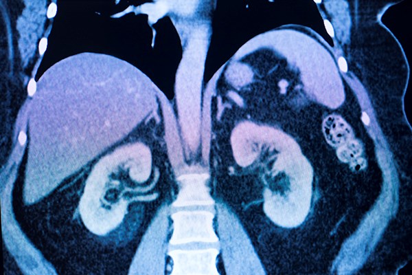
The degree to which contrast media is directly responsible for the development of acute kidney injury is hotly debated.
Contrast-induced nephropathy (CIN) is the development of acute kidney injury (AKI) as a direct result of the administration of iodinated contrast media (CM). This is a causative diagnosis with a direct cause-and-effect relationship.
Post-contrast AKI is a correlative diagnosis in that it is the development of AKI after administration of CM. This correlation does not equate to causation. The American College of Radiology (ACR) Manual on Contrast Media states that these terms are neither synonymous nor interchangeable.1
Past studies have not used a standard diagnostic criterion for CIN and PC-AKI. Most use both an absolute and relative increase in serum creatinine levels to make the diagnosis. According to the Acute Kidney Injury Network (AKIN), AKI is present when there is an increase in serum creatinine of more than 0.3mg/dL, an increase of more than 50% from the patient’s baseline creatinine level, or the presence of oliguria (< 0.5 mL/hr urine output) for more than 6 hours within 48 hours.2
Intravascular CM are concentrated solutions containing monomeric or dimeric tri-iodobenzene with differing side chains. Iodine, with a high atomic number (Z=53), imparts an increased density to the solution, which allows for visual contrast versus anatomic structures.3 CM can be classified as ionic or nonionic as well as high-, low-, and iso-osmolar. First generation CM were high osmolar agents (> 1200 mosm/kg) compared to plasma osmolarity of 280-290 mosm/kg. Low osmolar contrast media (LOCM) and iso-osmolar contrast media (IOCM) have largely replaced the use of high osmolar contrast media (HOCM). The mechanism by which CIN is thought to occur is due to 3 potential causes:
- Medullary ischemia
- The creation of reactive oxygen species
- Direct tubular cell toxicity4
The degree to which CM is directly responsible for the development of AKI is a matter of considerable debate. Published studies measuring the incidence of CIN are composed entirely of observational trials, and recent studies suggest that the risk of CIN is overestimated. A randomized control trial (RCT) would provide the best level of evidence to determine whether there is a causal relationship between contrast and AKI, but designing such an RCT would not be possible given the ethical issues associated with such a trial. Consequently, there have not been any RCTs to date evaluating the risk of developing CIN.
The Evidence
The first report of CIN was published in 1954 describing the case of a 69-year-old male, posthumously diagnosed with multiple myeloma, who underwent intravenous pyelography and subsequently developed anuria.5 Older studies of CIN were performed when most contrast imaging studies utilized HOCM. Some conclusions regarding the association of CM with the development of AKI were extrapolated from patients who underwent cardiac angiography procedures, which greatly overestimates its risk compared to that of intravenous administration commonly encountered in emergency department settings.6 Calculating the risk of CIN is difficult to determine due also to a lack of standard-ization used in prior studies regarding the definition of CIN.
A recent meta-analysis found 28 studies, all observational, with many using an absolute rise in serum creatinine of 0.3 to 0.5 mg/dL or a relative increase of 25% from baseline within three days of contrast administration.7 A common limitation among these observational studies is that without randomization, there may be confounding variables that influence the selection of patients who receive IV contrast and those who do not. This has been addressed with the use of propensity scoring, which takes into account the likelihood that a patient would be assigned to either group based on known confounders that can cause AKI other than CM.6
There have been multiple single-center, retrospective comparisons of contrast-exposed versus contrast-unexposed patients that have failed to demonstrate a statistically significant increase in the risk of developing AKI after exposure to CM.8,9 A subgroup analysis in a study by Davenport et al. did find an association of CM with AKI in patients having an elevated baseline creatinine of ≥1.6 mg/dL;10 however, a recently published study by Hinson et al. did not find this same association.11
The meta-analysis by Aycock et al. included more than 100,000 patients from 28 observational studies. They found the risk of AKI from contrast-enhanced CT compared to non-contrast CT was not increased (odds ratio [OR] 0.94; 95% confidence interval [CI] 0.83 to 1.07). No risk was also seen in the 6 studies that used matching techniques (OR 0.98; 95% CI 0.92 to 1.05).7
Conclusion
Despite being widely feared by the medical community for decades, the risk of CIN has been seriously challenged by recent studies. Ultimately, RCT-based evidence is necessary to reveal an accurate incidence of CIN as well as to elucidate whether causality is present.
Previous observational studies, though limited by the effect of potential confounding, strongly suggest that the risk of CIN, at the very least, has been highly overestimated. The impact this common-belief has on physician diagnostic behavior has not been quantified.
Given the importance that CM has in the diagnosis of multiple life-threatening diseases, it is essential that policies and guidelines provide a realistic and evidence-based calculation of the risk of AKI due to use of these agents.
References
1. ACR Committee on Drugs and Contrast Media. ACR manual on contrast media (10.3 ed.). American College of Radiology; 2017.
2. Mehta RL, Kellum JA, Shah SV, et al. Acute Kidney Injury Network: report of an initiative to improve outcomes in acute kidney injury. Critical Care. 2007;11:R31.
3. Cormode DP, Naha PC, Fayad ZA. Nanoparticle contrast agents for computed tomography: a focus on micelles. Contrast Media Mol Imaging. 2014;9(1):37-52.
4. Geenen RWF, Kingma HJ, van der Molen AJ. Contrast-induced nephropathy: pharmacology, pathophysiology and prevention. Insights Imaging. 2013;4(6):811-820.
5. Bartels ED, Brun GC, Gammeltoft A, Gjørup PA. Acute anuria following intravenous pyelography in a patient with myelomatosis. Acta Medica Scandinavica. 1954;150(4):297-302.
6. Luk L, Steinman J, Newhouse JH. Intravenous Contrast-Induced Nephropathy-The Rise and Fall of a Threatening Idea. Adv Chronic Kidney Dis. 2017;24(3):169-175.
7. Aycock RD, Westafer LM, Boxen JL, Majlesi N, Schoenfeld EM, Bannuru RR. Acute Kidney Injury After Computed Tomography: A Meta-analysis. Ann Emerg Med. 2018;71(1):44-53.e44.
8. Sinert R, Brandler ES, Subramanian RA, Miller AC. Does the Current Definition of Contrast-induced Acute Kidney Injury Reflect a True Clinical Entity? Acad Emerg Med. 2012;19(11):1261-1267.
9. McDonald RJ, McDonald JS, Carter RE, et al. Intravenous contrast material exposure is not an independent risk factor for dialysis or mortality. Radiology. 2014;273(3):714-725.
10. Davenport MS, Khalatbari S, Dillman JR, Cohan RH, Caoili EM, Ellis JH. Contrast material-induced nephrotoxicity and intravenous low-osmolality iodinated contrast material. Radiology. 2013;267(1):94-105.
11. Hinson JS, Ehmann MR, Fine DM, et al. Risk of Acute Kidney Injury After Intravenous Contrast Media Administration. Ann Emerg Med. 2017;69(5):577–586.e4.


