


Erythema multiforme is an acute skin disorder. While most cases are the result of an infection, the condition can also result from autoimmune disorders, malignancy, and drug use.
Case
A 24-year-old male presents to the emergency department with complaints of bilateral eye discharge and rash. He states the eye discharge had started unilaterally 10 days prior but then spread to involve both eyes. Six days prior he developed a cough for which he was prescribed azithromycin from a different emergency department, and began his azithromycin this morning. By afternoon he noted a rash on his body starting in the extremities, which rapidly progressed to include his mouth and lips. He has a history of seizures, but has not been on any antiepileptic medications since he was a child. He is unsure if he has had azithromycin in the past. He endorses diarrhea, mild odynophagia, and vague generalized abdominal pain. He denies any emesis or shortness of breath. He admits to smoking marijuana, but no other drug use.
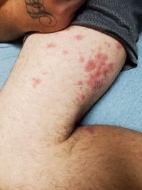
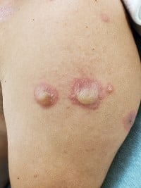
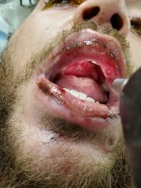
Physical exam illustrates a generally uncomfortable appearing male. Vitals noted tachycardia of 124, temperature of 98.6° F, RR 20, BP 126/84, and oxygen saturation 98% on room air. His eye exam was remarkable for bilateral conjunctival injection with a significant amount of thick white purulent discharge. Fluorescein stain showed no uptake. Oral exam demonstrated hard palate ulcerations, lips with significant erythema and blistering, and a thrush-like appearance of the tongue. Lung exam had diminished breath sounds bilateral, and the patient was continuously coughing up thick white mucus with a partially filled basin next to him. Skin exam demonstrated plaque-like targetoid rash noted over upper and lower extremities bilaterally, as well as on the lower abdomen. The chest, back, and upper extremities bilaterally also had blistering, large bullae, and sloughing of skin noted. There was no involvement of the palms, soles, or genital region.
Workup in the emergency department included chest xray, EKG, and labs including CMP, CBC, INR, urine, lipase, lactic, and troponin. There were no significant abnormalities noted. Urine drug screen was noted positive for cocaine and THC. Infectious workup was negative for COVID-19, flu, rapid strep, syphilis, and HIV. The patient was admitted with a clinical diagnosis of erythema multiforme.
Pathophysiology
Erythema multiforme (EM) is an acute skin disorder. The typical presentation is a target-like rash. It can also have vesiculobullous lesions. EM can be categorized into two groups: EM minor and EM major. EM minor has the typical target appearing rash; there may be bullae, none to minimal mucosal involvement, and no systemic symptoms. EM major has more extensive rash, mucosal involvement of at least two regions, and possible systemic involvement.1,2
The most common causes, about 90%, are thought to be infectious, with herpes simplex virus (HSV) and mycoplasma being particularly common. Only a small number of cases are thought to be drug-induced. Other causes include autoimmune, malignancy, and radiation.2
EM was once thought to be on the same spectrum as Stevens-Johnson syndrome (SJS), but is now considered a separate disease state.
Presentation
The rash is often an erythematous targetoid lesion, which begins on the extremities and spreads towards the trunk. It can also involve the soles and palms. The rash is typically not pruritic but can be in some cases. Mucosal involvement can include the mouth, genital region, and eyes. These can present as bullae or erosions which may be painful. Ocular involvement can present with discharge and can lead to conjunctival scarring and vision loss. Systemic symptoms can include malaise, arthralgia, cough, and dyspnea.2
The diagnosis is primarily clinical. Punch biopsy can be used to confirm. Biopsy should be performed in the center of the lesion. This will show keratinocyte necrosis, lymphocytic infiltrate in the superficial dermis, and epithelial intercellular edema.1,2
Management
Most EM will be self-limited, with resolution anywhere from 2-6 weeks. In patients with EM secondary to HSV, antivirals are typically not utilized in the acute presentation. They can be considered to prevent a recurrence. In EM secondary to medication exposure, the medication should be stopped.1,3
Systemic or topical steroids can be used for skin discomfort as well as oral lesions. Viscous lidocaine may also help with painful oral lesions. For severe oral lesions, patients may be unable to tolerate PO and require parenteral nutrition. Topical steroid drops and lubricants can be used for ocular involvement. An ophthalmologist should always be consulted if there is ocular involvement due to the potential for vision loss.
Disposition depends on case severity. In cases with dehydration due to decreased intake, inadequate pain control, significant systemic involvement, or secondary infection, consider admission. Some patients may require ICU admission and management similar to burn patients. Less involvement can be discharged home and follow up with dermatology outpatient.
Case Conclusion
Through his hospital stay, he was evaluated by infectious disease, dermatology, and ophthalmology.
Further lab work on admission was negative for HSV, hepatitis panel, blood cultures, and Epstein Barr virus. C-reactive protein was elevated to 123 (ref 0-10). His ANA, cold agglutinin screen, and rheumatoid factor were all negative. His Mycoplasma pneumoniae antibody IgG was positive; however, his Mycoplasma pneumoniae antibody IgM was negative, indicating past exposure but no current infection.
Ophthalmology placed him on oral doxycycline, topical erythromycin, and topical gatifloxacin drops.
Dermatology began him on hydrocortisone 100 mg every 8 hours, plus topical steroids. Skin biopsy illustrated detachment of the epidermis from the underlying dermis, full-thickness necrosis of the epidermis, and mild perivascular superficial lymphohistiocytic infiltrate. All of which are seen in EM. The presentation of acral to central spread of rash and generally nontoxic appearance of the patient supported EM over SJS.
He had clinical improvement while in the hospital. He completed a 7-day course of oral doxycycline and erythromycin eye drops. He was discharged on topical steroids, gatifloxacin eye drops, prednisolone eye drops, and a 2-week course of oral prednisone. His discharge diagnosis was EM with mucocutaneous involvement.
In this patient, many causes of EM were considered. He was negative for many viral and bacterial sources of infection. Autoimmune workup was also normal. He did have the recent use of a macrolide antibiotic. He also admitted to frequent nasal insufflation of cocaine after the results of his urine drug screen came back positive. There have been several case reports where azithromycin has been implicated in both EM and SJS.4,5 However, there is also a consideration of a contaminant in his cocaine resulting in his symptoms.
References
- Hafsi W, Badri T. Erythema Multiforme. [Updated 2020 Aug 15]. In: StatPearls [Internet]. Treasure Island (FL): StatPearls Publishing; 2021 Jan-. Available from: https://www.ncbi.nlm.nih.gov/books/NBK470259/
- Wetter DA. Erythema multiforme: Pathogenesis, clinical features, and diagnosis. March 2021. https://www.uptodate.com/contents/erythema-multiforme-pathogenesis-clinical-features-and-diagnosis. Accessed May 10, 2021.
- Wetter DA. Erythema multiforme: Management. November 2019. https://www.uptodate.com/contents/erythema-multiforme-management?search=erythema%20multiforme%20treatment&source=search_result&selectedTitle=1~150&usage_type=default&display_rank=1#H1. Accessed May 10, 2021.
- Fan Xiaomei, Luo Yong, Lu Jieluan, Xu Jinji, Chen Qing, Guo Huijuan, Jin Ping. Erythema Multiforme Major Associated With Community-Acquired Pneumonia: Lessons From a Case Report. Front. Pediatr., 29 July 2021. https://www.frontiersin.org/articles/10.3389/fped.2021.698261/full
- Brkljacić N, Gracin S, Prkacin I, Sabljar-Matovinović M, Mrzljak A, Nemet Z. Stevens-Johnson syndrome as an unusual adverse effect of azithromycin. Acta Dermatovenerol Croat. 2006;14(1):40-5. PMID: 16603101.