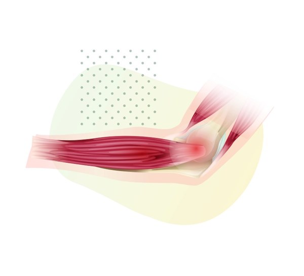
Morel-Lavallée lesions (MLLs) are soft tissue internal degloving injuries that develop following high-velocity trauma, most commonly, motor vehicle collisions.
Impact transmitted into the soft tissue causes a shearing force that separates the hypodermis from the underlying fascia, resulting in the disruption of lymphatics and capillary beds, and ultimately the development of a fluid collection. This fluid collection becomes necrotic and elicits a chronic inflammatory response.1
Because they often present several days following the traumatic impact, MLLs often go undiagnosed or have a delayed diagnosis, which impedes prompt treatment. This can lead to the development of a chronic lesion and increases the risk of complications including life-threatening infections. Thus, it is important to recognize the signs of MLLs, which include swelling, ecchymosis, fluctuance, and hypermobility of the skin.2 MLLs are usually associated with fractures of the greater trochanter, femur, or pelvis, but have been reported in the knee, gluteal region, and abdomen, with few reports in the literature of upper extremity MLLs.1
CASE REPORT
A 42-year-old female with no pertinent medical history presented to the emergency department with a chief complaint of 1 day of left forearm pain. The patient stated that she was on a boat, when a rope became wrapped around her forearm, and then pulled with extreme force, causing a soft tissue twisting injury. The patient initially noted some swelling and erythema, and tried over the counter analgesics, but continued to experience progressive swelling and discomfort. At the time of presentation, the patient was complaining of pain radiating to her upper arm and into her hand. She denied numbness, tingling, or any other symptoms.
The patient was a well-appearing middle-aged female in no acute distress. Vital signs demonstrated a blood pressure of 111/67, heart rate of 89 bpm, respiratory rate of 16, oxygen saturation of 98%, and temperature of 36.9°C. The patient rated her pain level as a 5 on a scale of 1-10.
On physical exam, there was an obvious deformity and pain to palpation of the left upper extremity near the proximal left wrist and distal forearm. Range of motion was limited in wrist flexion and extension as well as elbow flexion and extension secondary to pain. Patient had an 18 cm x 13 cm area of swelling and erythema to the volar aspect of the left forearm with cutaneous abrasions from the rope. Compartments were soft, and radial pulses were 2+ bilaterally. No crepitus was appreciated. Sensation was intact. The extremity was warm, with a capillary refill of less than 2 seconds. There was normal range of motion of the shoulder, hand, and fingers.
A two-view X-ray of the left forearm and a two-view X-ray of the left wrist were obtained to evaluate for an acute fracture or dislocation. No acute fractures or dislocations were observed, joint spaces were preserved, and alignment was maintained. Soft tissues appeared normal.
The patient was given oral narcotics for pain.
On reassessment, the patient continued to have pain out of proportion to the exam. Therefore, a CT scan of the left upper extremity with IV contrast was ordered to assess for further injury. The CT scan described soft tissue and subcutaneous fat edema with a small amount of fluid along the palmar aspect of the forearm measuring up to seven mm in thickness. No vascular injury was detected.
Blood work was obtained to evaluate for evidence of rhabdomyolysis or acute kidney injury. Total creatine kinase was slightly higher than the reference range at 587 U/L (normal 30-135 U/L). Basic metabolic panel revealed no electrolyte abnormalities and normal creatinine and GFR. The patient continued to have pain in the emergency department and was given IV narcotics for pain.
Based on the CT scan findings as well as the mechanism of injury, the patient was diagnosed with a Morel-Lavallée lesion. The surgical team was consulted and evaluated the patient. They agreed with the diagnosis and admitted the patient for further evaluation and treatment.
Treatment consisted of pain management and an evaluation by hand surgery, who recommended operative intervention. The patient went to the OR the next day for a left forearm exploration, washout, fasciotomy, and incision and drainage with packing. The patient received antibiotics prior to surgery. Intraoperative wound culture did not grow any organisms.
Prior to discharge, the patient met all of physical therapy’s mobilization requirements and was taught how to perform wound care. The patient was discharged from the hospital on day three with oral antibiotics, pain medications, and a follow-up appointment in the hand surgery clinic within 1 week.
DISCUSSION
This case is of interest as it highlights that Morel-Lavallée lesions can be found in the upper extremity and should be considered when the mechanism of injury involves a shearing force. This patient’s initial presentation suggested that she had a fracture of either her radius or ulna secondary to the swelling and obvious deformity of her forearm. Therefore, an X-ray was done first to evaluate.
Once the patient’s x-ray came back negative for fracture, suspicion was higher for other underlying pathology given the degree of swelling and pain. Although in this case the patient had no fracture, it is important to note that the patient could still have a MLL in concurrence with a fracture. Literature review shows that MLLs have been diagnosed in both traumas with and without fractures.3 Had this patient presented with a fracture, it is possible that her swelling and deformity would have been attributed to a fracture rather than an MLL.
Missed or delayed diagnosis of this MLL could have led to complications and permanent contour deformity. It is important to note that MLLs are underreported and are often misdiagnosed, therefore the published rate of incidence is most likely an underestimation. This makes it difficult to establish an overall incidence, as well as the rate of occurrence in tandem with fractures.
A thorough literature review was conducted using the following keywords: Morel-Lavallée lesion, emergency department, upper extremity, internal degloving, degloving injuries, upper, arm, shoulder, chest, abdominal, neck, head, spine, spinal, wrist, or hand. This resulted in 21 articles being found. Of these, 3 were in reference to case reports of Morel-Lavallée lesions in the upper extremity. Two of these cases were diagnosed outside of the emergency department. One report was diagnosed 10 months after injury during a follow up with hand surgery.4 The second report was diagnosed during follow-up with orthopedic surgery after an ED visit.3 It is unclear how the third report was diagnosed, as it is a radiology case report.5
Our case report is unique, as it was an upper extremity MLL diagnosed by emergency physicians during the patient’s first evaluation.
This resulted in prompt identification and treatment of the patient’s lesion, minimizing chances of infection and permanent deformity. Furthermore, this patient was promptly seen by the hand surgeons on call and was taken to the OR. Clinical course was uncomplicated. Ramaseshan et al. reported about 33-44% of Morel-Lavallée lesions are misdiagnosed.6 This rate of misdiagnosis is suspected to be higher in upper extremity lesions, as they are quite rare and therefore may not be in a physician’s primary differential. Vanhegan et al. reviewed 204 Morel-Lavallée cases in 29 published papers and found the incidence of Morel-Lavallée lesions based on location was reported to be the greater trochanter or hip 30.4%, thigh 20.1%, pelvis 18.6%, knee 15.7%, gluteal region 6.4%, lumbosacral 3.4%, abdominal wall 1.5%, calf or lower leg 1.5%, head 0.5%, and unspecified 2.0%.7 Given the distribution reported above, it can be deduced that the unspecified reports may encompass the upper extremity MLLs.
Morel-Lavallée lesion presentations differ between patients and cases. Presentations may also differ based on the chronicity of the lesion. They can present as painful lesions or even nonpainful lesions secondary to hypoesthesia from damage to cutaneous nerve branches.8 They can mimic various disease processes such as contusions, DVTs, cellulitis, fractures, soft tissue injuries, or abscesses.
It is important to consider Morel-Lavallée lesions when working up patients with traumatic injuries, as misdiagnosis may lead to complications such as infection, necrosis, permanent deformity and even death. There is a reported 1.7% comorbidity rate.9 Morel-Lavallée lesions have also been reported to cause hemorrhagic shock and death secondary to active bleeding into the lesion.8
Gold standard for diagnosing Morel-Lavallée lesions is MRI; however, CT scans and even point of care ultrasound have been found to be useful.10 Early detection can alter the management of MLLs and positively change clinical outcomes, thus it is important for emergency physicians to consider MLLs when evaluating and treating traumatic injuries.
CONCLUSION
Although MLLs are relatively uncommon, this may be confounded by the number of MLLs going undiagnosed. Thus, early recognition of MLLs is imperative to ensure prompt treatment.
If left untreated, MLLs can lead to chronic and debilitating sequelae including infection, skin necrosis, and impaired function and mobility.11
Furthermore, it is important for emergency physicians to maintain a high index of suspicion for MLLs in cases involving blunt trauma, even when the injury is in locations that are uncharacteristic of MLLs, such as the upper extremities and in cases where fractures are not present.
References
- Diviti S, Gupta N, Hooda K, Sharma K, Lo L. Morel-Lavallee Lesions-Review of Pathophysiology, Clinical Findings, Imaging Findings and Management. J Clin Diagn Res. 2017;11(4):TE01-TE04.
- Scolaro JA, Chao T, Zamorano DP. The Morel-Lavallée Lesion: Diagnosis and Management. J Am Acad Orthop Surg. 2016;24(10):667-672.
- Ab Halim MAH, Rampal S, Devaraj NK, Badr IT. A peculiar case of Morel-Lavelle lesion of upper limb. Med J Malaysia. 2020;75(5):594-596.
- Cochran GK, Hanna KH. Morel-Lavallee Lesion in the Upper Extremity. Hand (NY). 2016;12(1):NP10–NP13.
- Padmanabhan E, Rudrappa RK, Bhavishya T, Rajakumar S, Selvakkalanjiyam S. Morel-Lavallee Lesion: Case Report with Review of Literature. J Clin Diagn Res. 2017;11(7):TD05-TD07.
- Ramaseshan K, Bauler LD, Mastenbrook J. Morel-Lavallée Lesion of the Anterior Leg: A Rare Anatomical Presentation. BMJ Case Rep. 2020;13(2):e233295.
- Vanhegan IS, Dala-Ali B, Verhelst L, Mallucci P, Haddad FS. The morel-lavallée lesion as a rare differential diagnosis for recalcitrant bursitis of the knee: case report and literature review. Case Rep Orthop. 2012;2012:593193.
- Claassen L, Franssen MA, de Loos ER. A Rare Case of Hemorrhagic Shock: Morel-Lavallée Lesion. Clin Pract Cases Emerg Med. 2019;3(4):417-420.
- Kim WJ, Lee HS, Won SH, Hong YC, Lee DW, Lee JH, Kim CH. A Morel-Lavallée Lesion of the Proximal Calf in a Young Trauma Patient. Medicine (Baltimore). 2018;97(41):e12761.
- Annison DR, Smith M. Identification and triage of a Morel-Lavallée lesion using point of care ultrasound. Ultrasound. 2022;30(1):85-89.
- Molina BJ, Ghazoul EN, Janis JE. Practical Review of the Comprehensive Management of Morel-Lavallée Lesions. Plast Reconstr Surg Glob Open. 2021;9(10):e3850.


