Article: Cohen AL, Li T, Becker LB, et. al. Critical Care Alert: Femoral artery Doppler ultrasound is more accurate than manual palpation for pulse detection in cardiac arrest. Resuscitation. 2022;173:156-165.
Background
When performing cardiopulmonary resuscitation (CPR) on a patient, the standardized order is to determine if the rhythm is shockable, followed by two minutes of chest compressions, then a pulse check. However, manual palpation of the carotid artery pulse has an accuracy that ranges from 63 to 94%. This variability is due to factors such as body habitus, environmental stress, time limitations, and assessor experience.1 Despite this, manual palpation of the pulse is the primary mode for detection of return of spontaneous circulation (ROSC).
Objective
The primary goal of this study was to assess the accuracy of femoral artery Doppler ultrasound compared to manual palpation to detect a pulse in patients in cardiac arrest. The secondary goal was to determine whether peak systolic velocity (PSV) on Doppler ultrasound can accurately detect a pulse with an adequate blood pressure needed for perfusion, defined as SBP ≥ 60 mmHg.2
Design
The study was a prospective, cross-sectional, partially blinded, diagnostic accuracy study performed in a quaternary care Emergency Department (ED) between June 2019 to July 2021.
Methods
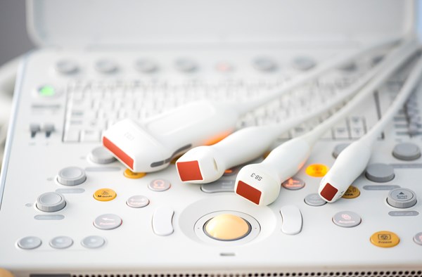
The study included adult patients ≥ 18 years old with nontraumatic cardiac arrest (both out-of-hospital and in-hospital) who had a femoral arterial line and a Doppler ultrasound-trained Emergency Medicine attending physician was available from the research team. Patients were excluded if they were a candidate for extracorporeal membrane oxygenation (ECMO). The research team underwent training for Doppler ultrasound for this study. The treatment team was blinded to the Doppler ultrasound exam. However, they were not blinded to arterial line blood pressure waveforms and measurements. The Doppler ultrasound was performed by a linear transducer probe in the vascular mode setting by the treatment team.
Prior to a pulse check, the ultrasound machine volume was turned off. During a pulse check, research personnel confirmed and recorded whether the treating team palpated a pulse, the presence or absence of Doppler ultrasound and arterial line waveforms, the highest PSV on Doppler ultrasound, and the highest SBP on the arterial line. Repeated measurements were recorded until ROSC was achieved or time of death. The enrolling ultrasonographer and a second Doppler ultrasound-trained sonographer, who was blinded to any patient information, reviewed all stored Doppler ultrasound images to confirm whether a Doppler waveform was present and the measurement of the highest PSV during the pulse check.
Outcomes
A receiver operator characteristic (ROC) curve analysis was performed to determine the cut-off value of PSV on Doppler ultrasound that is associated with SBP ≥ 60 mmHG. Accuracy, sensitivity, and specificity of Doppler ultrasound and manual palpation for detection of any pulse and a pulse with a SBP ≥ 60 mmHg were calculated and compared with McNemar’s test.
It was determined that a sample size of 308 pulse checks would be needed to estimate the accuracy of Doppler ultrasound for pulse detection with a width of 10% if the sensitivity of manual palpation was 80%, specificity was 70%, and the prevalence of a pulse was 20%. The study was stopped early after an interim analysis found the sensitivity of manual palpation was lower and the prevalence of a pulse was higher than predicted.
54 patients and 213 pulse checks were included for the primary outcome detection of any pulse, with a median of 3 pulse checks per patient (interquartile range: 2–6 pulse checks). The median age was 81 years (IQR 66–87 years), predominantly male (67%), and white (63%), largely OHCA (61%), and predominantly non-shockable; asystole representing 28% and PEA 35% of initial ED rhythms.
Key Results
Doppler ultrasound had higher accuracy than manual palpation for detection of any pulse (95.3% vs. 54.0%; p < 0.001). When SBP ≥ 60 mmHg, the accuracy of Doppler ultrasound was lower, however still more accurate than manual palpation (77.6% vs. 66.2%; p = 0.011). For detection of a pulse with SBP ≥ 60 mmHg, the sensitivity of manual palpation was low at 47.4%; however, the specificity of manual palpation was higher than Doppler ultrasound (82.3% vs. 58.4%; p < 0.001).
210 pulse checks with PSV on Doppler ultrasound and arterial line SBP were included. The optimal cut-off value for PSV associated with a SBP ≥ 60 mmHg was 20 cm/s. To detect SBP ≥ 60 mmHg, the accuracy of PSV ≥ 20 cm/s on Doppler ultrasound was higher than manual palpation (91.4% vs. 66.2%; p < 0.001).
Limitations
The study was a single-center study with a small number of patients, repeated measurements for each patient were used. Treatment teams were not blinded to the arterial line. Carotid artery palpation has recently been found to be more sensitive than femoral artery palpation during cardiac arrest.3 However, in this study, the location of manual pulse detection (carotid or femoral) was not consistently recorded, as the carotid and femoral pulses were often simultaneously checked.
EM Takeaways
In this study, femoral artery Doppler ultrasound was significantly more accurate than manual palpation for detecting any pulse. A PSV ≥ 20 cm/st was found to predict a pulse with a SBP ≥ 60 mmHg, which was considered adequate for perfusion. In settings where Doppler ultrasound is available, femoral artery Doppler ultrasound is an accurate and objective tool to determine the presence of a pulse in cardiac arrest. External validation of this study, particularly the PSV cut-off on Doppler ultrasound, is needed before widespread adoption.
References
- Nakagawa Y, Inokuchi S, Morita S, et al. Accuracy of pulse checks in terms of basic life support by lifesavers, as lay persons. Circ J. 2010; 74: 1895-1899.
- Deakin CD, Low JL. Accuracy of the advanced trauma life support guidelines for predicting systolic blood pressure using carotid, femoral, and radial pulses: observational study. BMJ. 2000; 321:673-674.
- Yılmaz G, Bol O. Comparison of femoral and carotid arteries in terms of pulse check in cardiopulmonary resuscitation: A prospective observational study. Resuscitation. 2021; 162 56-62.


