


For new sonographers, looking for the common bile duct (CBD) can feel like that old school game called “Where’s Waldo” from the 1990s, but fret not. The goal of this article is to equip you with a targeted approach when looking for the common bile duct.
Anatomy Review
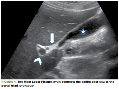
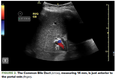
First, let’s review the landmarks that will orient us to where the CBD should be. As demonstrated in Figure A, the portal triad meets the gallbladder neck via the hyperechoic main lobar fissure. Notice in Figure B that the CBD is directly anterior to the portal vein. In the transverse view, it is typically lateral to the hepatic artery (though an estimated 15% of patients will have that orientation reversed). When in doubt, color flow is recommended to distinguish between the hepatic artery, which does demonstrate flow, and the CBD, which does not. This will also help you differentiate between the CBD and the IVC (Figure B).
HUNTING FOR THE CBD
General Approach
The curvilinear probe is the ideal choice for this exam type. However, if the CBD can only be identified through an intercostal window, the phased array probe can be effective. You should look for either the “exclamation point sign,” the “Mickey Mouse sign,” or the gallbladder by one of three methods: the subcostal sweep, the “X-7” approach, or the axillary transhepatic approach.
The subcostal sweep refers to sweeping beneath the right rib cage in a medial to lateral fashion. The X-7 approach places the probe approximately 7 cm to the right of the xiphoid process over the rib cage. The transhepatic axillary approach refers to scanning through the liver in the right axilla similar to the technique for the right upper quadrant view of a FAST exam.
ANATOMICAL LANDMARKS
Exclamation Point Sign
Sweep through the liver looking for the “exclamation point sign.” When visualizing the gallbladder in its long axis, the exclamation point is composed of two structures: the gallbladder and the portal vein. As demonstrated in figure C, the portal vein is the little “period,” and the gallbladder is the long structure towering over the period. With the portal vein identified, you can then apply your anatomy knowledge and find the CBD anterior to the portal vein. Remember you can also use color flow to verify that it is a duct rather than a vascular structure.
Mickey Mouse Sign
The “Mickey Mouse Sign” refers to the portal triad. As demonstrated in Figure Z, imagine that Mickey is facing you on the screen. In about 85% of patients, Mickey’s right ear is the CBD and his left ear is the hepatic artery. Color flow can be of great assistance in distinguishing the two structures.
Gallbladder
Identify the gallbladder in its long axis. Sweep through the gallbladder looking for its neck, then recall that the gallbladder neck sits near the liver’s main lobar fissure. Track the hyperechoic main lobar fissure in the opposite direction of the gallbladder, which should bring you to the portal triad.
Measuring CBDs
You found it! Now let’s assess it for dilation as a sign of biliary obstruction. The intraluminal diameter of the CBD is measured from the inner wall to the inner wall. As a general rule of thumb, the CBD should be less than the first digit of the patient's age1. For example, a 40-year-old should have a CBD < 4mm, while an 80-year-old should have a CBD < 8mm.
Still can't find it?
Consider the possibility that it is so dilated that you did not initially recognize it. Once you have identified the portal vein, look for the "double-barrel sign." Ultrasound is specific for finding CBD dilatation.
You still can't find it. What's next?
We advocate that assessing the CBD should be part of every biliary ultrasound exam. However, there is a paucity of evidence regarding the sensitivity of ultrasound for evaluating CBD. Furthermore, some research suggests that in the absence of abnormal biliary labs and other ultrasound signs of gallbladder pathology (ie, wall thickening, pericholecystic fluid, and gallstones), the identification of the CBD rarely changes management2.
Identifying the common bile duct can be difficult for new sonographers. By familiarizing yourself with the anatomy and practicing your technique, you can optimize the success of your hunt.
References
- Mike Mallin & Matthew Dawson. “Introduction to Bedside Ultrasound: Volume 2.” Emergency Ultrasound Solutions, 2013.
- Lahham S, Becker BA, Gari A, Bunch S, Alvarado M, Anderson CL, Viquez E, Spann SC, Fox JC. Utility of common bile duct measurement in ED point of care ultrasound: A prospective study. Am J Emerg Med. 2018 Jun;36(6):962-966. doi: 10.1016/j.ajem.2017.10.064. Epub 2017 Nov 20. PMID: 29162442.