


A 55-year-old female presents to the emergency department with 2 days of worsening right eye pain and conjunctival injection, now with proptosis (Figure 1). She had been seen the day prior and had been sent home with a presumptive diagnosis of conjunctivitis. Her eye examination is significant for mild proptosis, chemosis, and severe conjunctival injection of the right eye. Pupils are 4mm, equal, round, and reactive to light and accommodation. Visual acuity is 20/40 bilaterally without corrective lenses. Left eye is normal with no obvious gross defects. After reviewing her chart, you begin to wonder what else should be on your differential.
Background
The differential diagnosis for “red eye” can be quite broad (Table 1). Therefore, careful history and physical examination are of paramount importance. Specific factors to consider include the following: chronicity of illness, degree of pain, history of trauma or recent procedure, presence of visual changes, presence of proptosis, presence of fever, restriction of eye movement, cranial nerve involvement, and medical comorbidities.1
Because of this patient's unusual and repeat presentation, an orbital computerized tomography (CT) scan is performed, ultimately revealing the diagnosis of orbital pseudotumor, an idiopathic non-neoplastic inflammatory syndrome of the orbit.2, 3
Clinical Features and Diagnosis
Idiopathic orbital inflammation, often called orbital pseudotumor or orbital inflammatory syndrome, can affect every structure in the orbit, including the lacrimal gland, extraocular muscles, orbital fat, and the optic nerve.4 Orbital pseudotumor typically affects patients less than 50 years old and may occur unilaterally or bilaterally. It is a common cause of unilateral exophthalmos (as opposed to Graves' disease, which is most often bilateral). Bilateral involvement occurs in less than 10% of cases.2
Typical presenting symptoms include pain, erythema, swelling, proptosis, and restrictions in eye movement.2- 6 The diagnosis can be made with careful patient history, ultrasonography, CT, and/or magnetic resonance imaging (MRI).2 Diffuse infiltration of the orbit, inflammation of the sclera, and/or optic nerve involvement may be seen.2 CT of the orbits will show involvement of the tendinous insertion of the extraocular muscles, which distinguishes it from thyroid associated orbitopathy, where the insertion point is spared (Figure 2).
Moreover, a CT will demonstrate thickening and fat stranding around the muscle belly (Figure 3). Diagnosis may be confirmed with a biopsy referred to as an orbitotomy.2 However, if suspicion is high enough, response to corticosteroids can also be used to confirm a diagnosis without the need for biopsy.2
Pathophysiology
Although the pathophysiology of this condition remains unknown, it has been hypothesized that immune-mediated processes are the most likely underlying mechanism.1 Infections and aberrant wound healing have been proposed as other etiologies. Several infectious diseases, such as viral upper respiratory infection, Borrelia burgdorferi, and Streptococcal pharyngitis have also been linked with orbital pseudotumors. Pathological findings of biopsies are often nonspecific, and may only reveal benign lymphoid hyperplasia and inflammatory cell infiltration with necrotizing vasculitis.2
Management
The cornerstone of treatment includes systemic corticosteroid therapy.3,8 It is reported that approximately 75% of patients have a remarkable improvement after 24-48 hours of treatment.2,7 Although most cases have a rapid and dramatic response to steroids, there is also a chronic form with progressive fibrosis associated with a mild or poor response to steroids.2,8 In such cases, chemotherapy or radiation therapy is utilized.2 Collectively, it has been found that almost 80% of patients have a positive outcome.2 However, relapses are common, especially in bilateral disease.2
It is important to have ophthalmology involved for treatment and follow-up of this condition as well as to consider ruling out other rare but severe causes of orbital inflammation, such as thyroid-associated orbitopathy, sarcoidosis, infectious orbital cellulitis, orbital myositis, orbital vasculitis, Sjogren's syndrome, Wegener's granulomatosis, or malignant ocular tumors.2
The patient was admitted to the medical floor for intravenous methylprednisolone, as nausea and vomiting prevented her from tolerating oral steroids. On examination by ophthalmology the following morning, her signs and symptoms were much improved. She was discharged on a prednisone taper.
TABLE 1. Differential Diagnosis
| Viral conjunctivitis |
| Bacterial conjunctivitis |
| Allergic conjunctivitis |
| Acute angle closure glaucoma |
| Subconjunctival hemorrhage |
| Keratitis |
| Iritis/Anterior uveitis/Iridocyclitis |
| Episcleritis |
| Scleritis |
| Endopthalmitis |
| Herpes zoster opthalmicus |
| Orbital/postseptal cellulitis |
| Preseptal cellulitis |
| Corneal ulcer |
| Retroorbital hematoma |
| Graves' ophthalmopathy |
| Orbital pseudotumor |
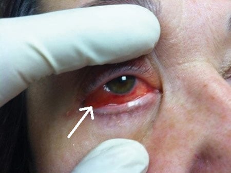 Figure 1. Conjunctival Injection and Proptosis
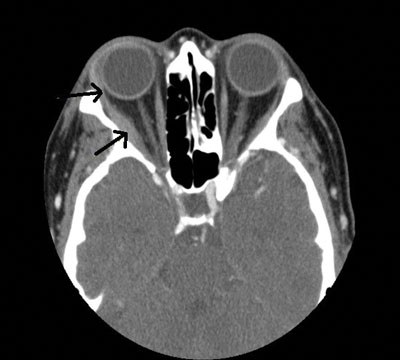 Figure 2. Axial CT of the Orbits
Shows thickening of the right lateral rectus muscle belly and its tendinous insertion (black arrows). Involvement of the tendinous insertion distinguishes it from thyroid associated orbitopathy, in which the insertion point is spared.
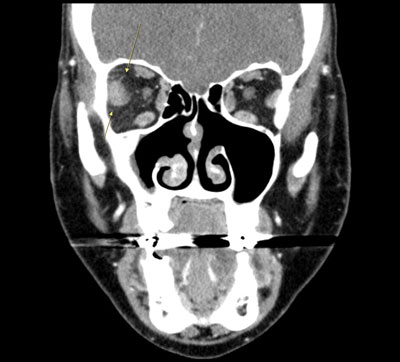 Figure 3. Coronal CT of the Orbits
Shows thickening and fat stranding around the right lateral rectus muscle belly (yellow arrows).
Conclusion
It is imperative to entertain a broad differential when encountering any ophthalmologic complaint. It is especially important when patients present for a return visit with new or changing symptoms. In general, the diagnosis of orbital pseudotumor should be entertained in any patient presenting with acute onset unilateral proptosis.
References
- Welsh L, Aaronson E, Eicken J, Geyer B, eds. EM Fundamentals: The Essential Handbook for Emergency Medicine Residents. Irving, Texas: EMRA; 2016.
- Chaudhry IA, Shamsi FA, Arat YO, et al. Orbital pseudotumor: Distinct diagnostic features and management. MEAJO. 2008;15:17-27.
- Finger PT for New York Eye Cancer Center. Orbital Pseudotumor. https://eyecancer.com/eye-cancer/conditions/orbital-tumors/orbital-pseudotumor/. Accessed August 3, 2015.
- Hong ES, Birkholz ES, Wendel L, Allen RC. Orbital inflammation secondary to bisphosphonate therapy (orbital pseudotumor). EyeRounds.org. January 4, 2010; Available from: http://www.EyeRounds.org/cases/101-orbitalpseudotumor.htm.
- Kapur R, Sepahdari A, Mafee M, et al. MR Imaging of Orbital Inflammatory Syndrome, Orbital Cellulitis, and Orbital Lymphoid Lesions: The Role of Diffusion-Weighted Imaging 2008. AJNR. 2009;30:64-70.
- Lusby F, Zieve D, Ogilvie I for U.S. National Library of Medicine. Orbital pseudotumor: MedlinePlus Medical Encyclopedia. https://www.nlm.nih.gov/medlineplus/ency/article/001623.htm. Accessed August 03, 2015.
- Mombaerts I, Goldschmeding R, Schlingemann RO, Koornneef L. What is orbital pseudotumor? Surv Ophthalmol. 1996;41(1):66-78.
- Yuen SJA, Rubin PA. Idiopathic Orbital Inflammation. Arch Ophthalmol. 2003;121(4):491-499.