


Blunt cardiac injury (BCI) is ill-defined but refers to injury to the myocardium from blunt chest trauma.
Unfortunately, there are no isolated tests one can perform to diagnose the condition; rather, the diagnosis relies on the overall clinical picture and clinical gestalt. While major complications are uncommon, BCI can be life-threatening and can result in ventricular wall rupture. More commonly, BCI manifests as clinically insignificant arrhythmias and mildly elevated troponins, indicating inconsequential myocyte injury.1
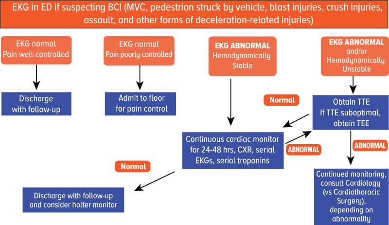
Mechanism of Injury
The incidence of BCI varies widely from 8%-76%, largely due to the lack of a specific definition or diagnostic tests.1 Bansal et al. purports that 25% of mortalities in traumatic injuries are correlated with BCI.2 BCI is most commonly seen in the setting of motor vehicle accidents.2 Pedestrians struck by a vehicle, blast injuries, crush injuries, assault, and other forms of deceleration-related injuries comprise the next most common causes. Anatomically, the two main mechanisms of injury include when the myocardium comes into contact with the surrounding bony structures (sternum, spine) or from shearing stress forces.3 The most commonly injured areas of the heart follow this order: right ventricle, right atrium, left ventricle, left atrium, atrial septum, valves (mitral and aortic > tricuspid and pulmonic), ventricular septum, and coronary arteries.4
Diagnostic Testing
The main diagnostic tools to assist with making this diagnosis include troponin/CK-MB, EKG, and echocardiography. In a retrospective study, Fegheli et al. demonstrated that troponin elevation was a sensitive test for diagnosis of BCI but was not specific in patients suffering blunt trauma for this injury.5
EKG is another useful test in diagnosing BCI, as 40-83% of patients with BCI will have EKG changes.6 Arrhythmias are the most commonly identified abnormality on EKG. Sinus tachycardia is the most commonly identified arrhythmia, though additional arrhythmias include ventricular fibrillation, atrial fibrillation, right bundle branch block, first degree heart block and third degree heart block. If the left ventricle is injured, common EKG findings include ST-T wave changes, diffuse ST-segment elevation, and Q waves.7 The degree of cardiac complications is not correlated with the degree of EKG changes, though, and special care must be made to bring together the whole clinical picture.1
Performing echocardiography is the next diagnostic study one can employ when assessing for presence and degree of BCI. While TOE and TEE are preferable to TTE, they require more resources, are more invasive and are not necessary in all patients. While echocardiography has not been shown correlate closely with EKG changes or troponin elevations, it is especially useful in that in can visualize cardiac structures, identify wall motion abnormalities, and better identify which patients may suffer from acutely clinically significant BCI.8
Management Algorithm
References
1. Blunt Cardiac Injury. Statpearls.com. https://statpearls.com/kb/viewarticle/18884/. Published 2019.
2. Bansal MK, Maraj S, Chewaproug D, Amanullah A. Myocardial contusion injury: redefining the diagnostic algorithm. Emerg Med J. 2005;22(7):465–469.
3. Tenzer ML. The spectrum of myocardial contusion: a review. J Trauma. 1985;25(7):620-627.
4. Pasquale M, Fabian TC. Practice management guidelines for trauma from the Eastern Association for the Surgery of Trauma. J Trauma. 1998;44(6): 941–956.
5. Fegheli NT, Prisant LM. Blunt myocardial injury. Chest. 1995;108(6):1673-1677.
6. Snow N. Richardson JD, Flint LM Jr. Myocardial Contusion: Implications for patients with multiple traumatic injuries. Surgery. 1982;92(4):744-750.
7. Braunwald E, ed. Braunwald’s Heart. 5th ed. Philadelphia: WB Saunders;1997.
8. Karalis DG, Victor MF, Davis GA, et al. The role of echocardiography in blunt chest trauma: a transthoracic and transesophageal echocardiographic study. J Trauma. 1994;36(1):53-58.