


A 67-year-old male with a past medical history of hypertension, poorly controlled Type 2 diabetes mellitus, obstructive sleep apnea, and a pacemaker presented to the ED with dry heaving and nausea for 5 days. He had not eaten for the past 3 days. He had been prescribed ondansetron and a BRAT diet by his primary care physician 2 days prior to arrival, with little relief. He denied fevers, chest pain, or dyspnea.
On presentation, his vital signs were normal. On physical examination, the patient had mild tenderness to palpation of the epigastric region, but no rebound or guarding.
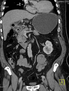
Abdominal labs along with a CT abdomen and pelvis with contrast were ordered. Morphine, ondansetron, and fluids were administered. Blood work was significant for leukocytosis to 21.2 and lactic acid of 2.8. CT scan demonstrated a gastric outlet obstruction secondary to a gallstone that had eroded through the gallbladder wall into the duodenum with markedly distended stomach and distal esophagus. There was an air-filled communication between the gall bladder and duodenum consistent with a choledochofistula (figure 1).
The attending radiologist remarked this was consistent with Bouveret syndrome, a finding he had never seen in his 35 years of work. Both general surgery and GI were consulted. The patient started antibiotic treatment with metronidazole and ceftriaxone. He had a nasogastric tube placed and was admitted to the surgical floor for GI endoscopic retrieval the following day.
Discussion
Gallstone-induced ileus is a rare complication of cholelithiasis. Gastric outlet obstruction caused from a gallstone is even rarer. Bouveret syndrome is named after the French physician Léon Bouveret (1850–1929), who reported the manifestation of gastric outlet obstruction caused by an impacted gallstone in 1896.1
Gallstones that are too large to pass through the cystic and common bile ducts can result in chronic irritation and inflammation of the gallbladder wall. This can lead to formation of a bilioenteric tract with gallstone migration into the upper gastrointestinal tract causing obstruction of the duodenum.
Classic gallstone ileus involving the terminal ileum complicates only 0.3%–0.5% of patients with cholelithiasis. Bouveret syndrome represents approximately 1%–3% of all gallstone ileus cases.2
Diagnosis
The typical presentation involves an older person with nausea, vomiting, and possible hematemesis from duodenal or celiac artery erosions. Up to 82% of patients have had a history of abdominal pain or biliary colic prior to presentation.3 Patients often have increased bilirubin and alkaline phosphatase.
Diagnosis is best achieved with CT scan, patient presentation, and the Rigler Triad: bowel obstruction, pneumobilia, and an ectopic gallstone.4 The mortality is between 12-30% due to patients’ advanced age, comorbidities, and high rate of surgical complications.5
Management
Due to the relative rarity of the condition, there are no standardized guidelines for management. Several endoscopic, laparoscopic, and open surgical techniques have been mentioned in management. Endoscopy is a useful initial diagnostic and possible therapeutic measure. Endoscopy facilitates mechanical lithotripsy, laser lithotripsy, and extracorporeal shock wave lithotripsy.
With large impacted gallstones (>4 cm), early surgical intervention should be considered for removal.6 Various open and laparoscopic surgical techniques have been used depending on location of the obstruction and presence of inflammation or ulceration. Examining the remaining small intestine is crucial to ensure intestinal gallstones broken up by lithotripsy do not cause future obstruction.7
Case Conclusion
The patient underwent upper endoscopy with GI the following day. The entire stomach was normal. A large (approximately 3 cm) stone was found in the duodenal bulb causing complete obstruction (figures 2 and 3). Initial attempts to remove the gallstone with snare, tripod, and roth net were unsuccessful. Electrohydraulic lithotripsy was then done to break down the stone. A jagwire was passed under fluoroscopic guidance into the distal duodenum and pyloric dilation was performed to remove the remaining stone parts. A 1.5 cm fistula was found in the duodenal bulb (figure 4). This was suspicious for cholecystoduodenal fistula. The patient had no complications and was discharged from the hospital the next day. He had a follow-up EGD 8 weeks later that was unremarkable. There was no indication for cholecystectomy, as the patient was asymptomatic and the fistulous tract was decompressed.
References
- Haddad FG, Wissam M, Liliane D. Bouveret's syndrome: Literature review. Cureus. 2018.
- Chen Y-F, Su Y-S, Chen F-L, Lee W-S. Bouveret syndrome complicating gastric outlet obstruction and acute cholecystitis. Journal of Microbiology, Immunology and Infection. 2020;53(5):823-825.
- Yale SH, Tekiner H, Yale ES. Learning about the diagnosis of Bouveret syndrome from Bouveret. Journal of Microbiology, Immunology and Infection. 2021;54(2):339-340.
- Brennan GB, Rosenberg RD, Arora S. Bouveret Syndrome. RadioGraphics. 2004;24(4):1171-1175.
- Abdallah MA, Atiq M, Kushnir V, et al. Management of Bouveret's syndrome. Journal of Digestive Diseases. 2019;20(4):215-219.
- Caldwell KM, Lee SJ, Leggett PL, Bajwa KS, Mehta SS, Shah SK. Bouveret syndrome: Current management strategies. Clinical and Experimental Gastroenterology. 2018;11:69-75.
- Haddad FG, Wissam M, Mansour J, Liliane D. From Bouveret’s syndrome to gallstone ileus: The journey of a migrating stone! Cureus. 2018.