


Stop us if you've heard this presentation before: leg/calf pain and new swelling. They could have just come back from a 16-hour trip by plane or car, might be taking birth control, smoking, or all/ none of the above. Regardless of the history, we see this presentation in the ED all the time, and the big can't-miss diagnosis is going to be: deep venous thrombosis (DVT)!
Scenario
You are in your ED and a patient comes in with the above presentation - next step? History!
Quick refresher on the Wells' Score for DVT:¹
- Active cancer, immobilization for more than 3 days or major surgery in the last 3 months, calf swelling of more than 3 cm compared to the other leg, non varicose veins, entire leg swelling, localized tenderness along the deep venous system, unilateral pitting edema, paralysis/recent immobilization of the lower extremity, previous DVT all earn points.
- A score of 0 or lower - unlikely to be a DVT
- 1-2 is moderate risk - D-Dimer is sufficient to rule out DVT
- 3 or higher - Likely DVT, should receive an Ultrasound.
So, this patient in front of you earns a 3 or higher and it is time to order that formal ultrasound… or maybe it is our chance to use our ultrasound skills and diagnose at bedside!
All of the governing bodies of ultrasound agree that the standard for suspected DVT is Ultrasound. However, given that there is a lack of a single protocol - now is our time to look in depth at the 3 point DVT study, which might save you and your patient time in diagnosing a blood clot in the ED.
First Step: Secure the right equipment²
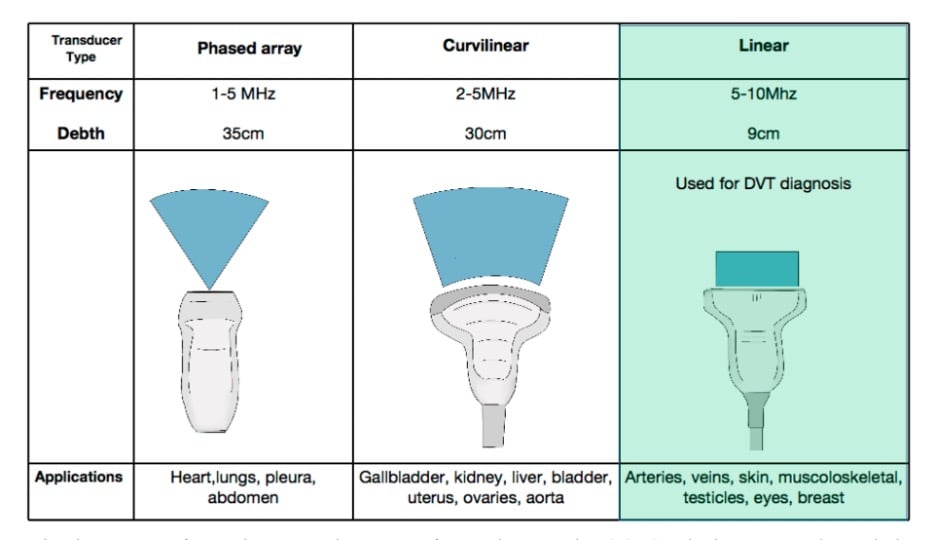
- With its higher frequency - images near the surface show higher resolution, making the linear probe ideal for this study.
Second Step: Placement of the patient and probe²
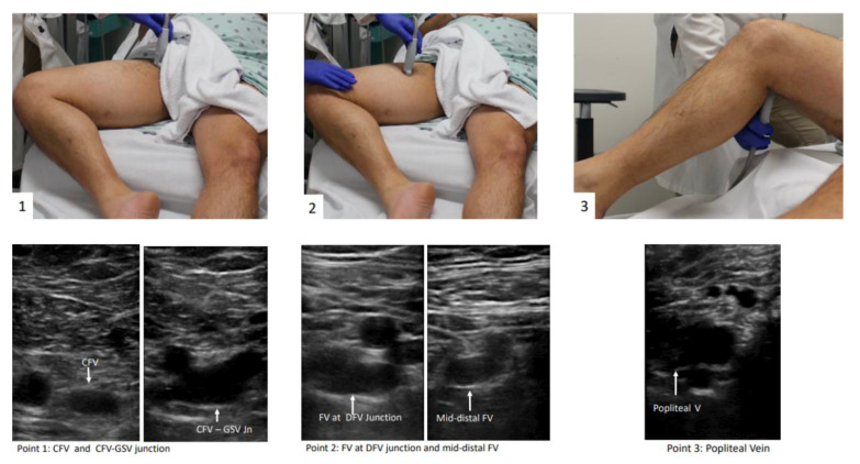
- Supine, with head elevated to 30° (can help with circulation and visualization)
- Externally rotate the hip + bend the knee = “Frog Leg”
- Common femoral vein = along the inguinal ligament in between the pubic symphysis and the anterior superior iliac spine. Remember VAN! - The vein should be medial to the artery.
- Superficial Femoral Vein = 1-2 cm down where the common bifurcates into deep and superficial.
- Popliteal vein = posterior crease of the knee and scan distal until it trifurcates
Third Step: Scanning²
- Inspect for thrombus, and if none are seen you will compress the vein in the transverse view.
- Apply enough pressure rapidly that the pulsatile artery compresses slightly
Example of echogenic clot distal femoral vein2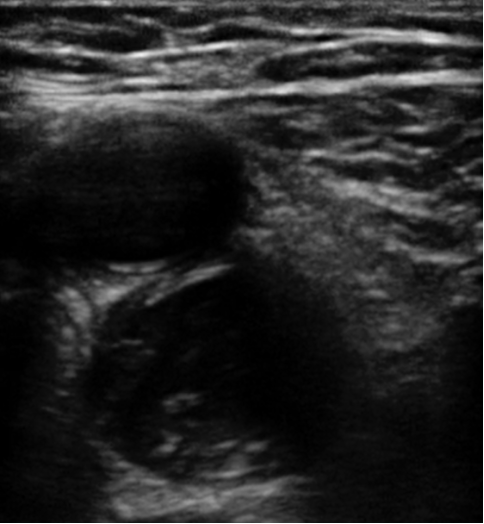
Example of non-compressibility in popliteal vein (top vessel)2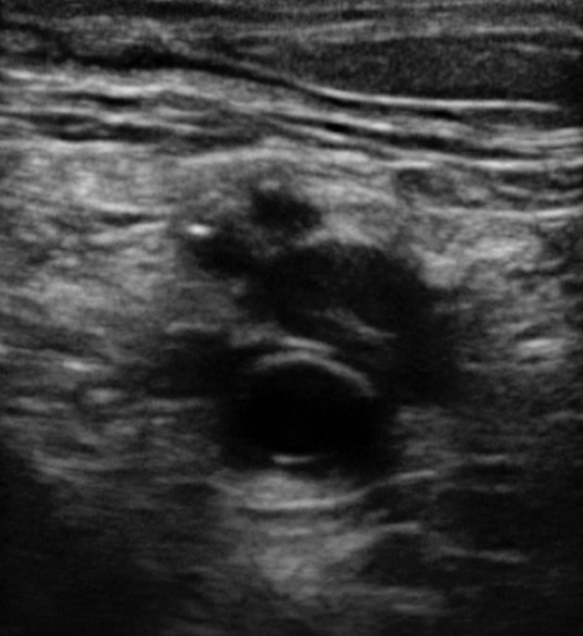
Sensitivity and Specificity
- Recent studies have shown good data supporting the sensitivity and specificity of DVT point of care ultrasound studies:
- HOCUS-POCUS (a prospective, multicenter trial on non-ICU hospitalized patients performed by hospitalists) showed a specificity of 95.7% and a sensitivity of 100%. Importantly it also shows that results saved about 5 hours over completed vascular results.⁷
- In 2009, the Journal of Thrombosis and Haemostasis looked at DVT evaluation - finding that at 3 months the rate of VTE was 2% in a rapid ultrasound vs. 1.2% in a complete vascular exam. Therefore, they determined that bedside ultrasound was a valid diagnostic test.⁸
- A 2019 meta-analysis pooling 17 studies in the emergency department, showed that both the 2-point and 3-point point of care studies had a sensitivity of 91, 90% and specificity of 98, 95%, respectively. Again, showing excellent diagnostic performance.⁹
References
- Needleman L, Cronan JJ, Lilly MP, et al. Ultrasound for Lower Extremity Deep Venous Thrombosis: Multidisciplinary Recommendations From the Society of Radiologists in Ultrasound Consensus Conference. Circulation. 2018;137(14):1505-1515.
- Varrias D, Palaiodimos L, Balasubramanian P, et al. The Use of Point-of-Care Ultrasound (POCUS) in the Diagnosis of Deep Vein Thrombosis. J Clin Med. 2021;10(17):3903.
- Troianos CA, Hartman GS, Glas KE, Skubas NJ, Eberhardt RT, Walker JD, Reeves ST. Guidelines for Performing Ultrasound Guided Vascular Cannulation: Recommendations of the American Society of Echocardiography and the Society of Cardiovascular Anesthesiologists. J Am Soc Echocardiogr. 2011;24(12):1291-1318.
- POCUS 101 Updates. Step-by-Step DVT Ultrasound Protocol: The 3-Point DVT Ultrasound Protocol. POCUS 101 by Vi Dinh, MD, FACEP, FDMS, RDCS.
- University Hospitals Emergency Medicine Residency. Intern Ultrasound of the Month: DVT Diagnosed at Bedside. The Land of EM blog. June 4, 2021.
- Greenwood-Ericksen M, Rempell J, Stone M. Ultrasound DVT Assessment. ALiEM Cards. Feb. 18, 2015.
- Fischer EA, Kinnear B, Sall D, et al. Hospitalist-Operated Compression Ultrasonography: a Point-of-Care Ultrasound Study (HOCUS-POCUS). J Gen Intern Med. 2019;34(10):2062-2067.
- Gibson NS, Schellong SM, Kheir DY, et al. Safety and sensitivity of two ultrasound strategies in patients with clinically suspected deep venous thrombosis: a prospective management study. J Thromb Haemost. 2009;7(12):2035-41.
- Lee JH, Lee SH, Yun SJ. Comparison of 2-point and 3-point point-of-care ultrasound techniques for deep vein thrombosis at the emergency department: A meta-analysis. Medicine (Baltimore). 2019;98(22):e15791.