


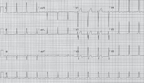
A 72-year-old male with a history of hyperlipidemia presents to the ED after an episode of exertional chest pain which has since resolved. What is your interpretation of his EKG?
Answer
This EKG shows a normal sinus rhythm with a ventricular rate of 82 bpm, normal axis, normal QRS duration, normal intervals, and biphasic T-waves in leads V2-V3. These findings, in the context of the patient’s history, are concerning for Wellens' syndrome.
Wellens' syndrome, also called "LAD coronary T-wave syndrome," describes an abnormal T-wave pattern seen in a pain-free state with recent history of anginal symptoms and is suggestive of a critical proximal LAD stenosis. It was first described in 1982 by Dutch cardiologist Dr. Hein J.J. Wellens, who found that 75% of patients with this pattern developed an extensive anterior STEMI if they did not receive coronary intervention.1
The pathophysiology of Wellens' syndrome is thought to involve a transient LAD occlusion which spontaneously resolves. Patients typically describe anginal pain which has resolved, and the characteristic EKG findings are seen in a pain-free state. These EKG findings represent coronary reperfusion, similar to what is seen after successful PCI.
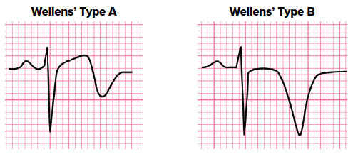
Wellens described 2 T-wave patterns: biphasic T-waves (type A), which are an early finding, and deeply inverted T-waves (type B), which are a later finding and more common (up to 75% of cases). The full diagnostic criteria include:
- Biphasic (type A) or deeply inverted (type B) T-waves in precordial leads, typically V2-V3
- Isoelectric or minimally elevated ST-segment (< 1 mm)
- No precordial Q-waves
- Preserved precordial R-wave progression
- Normal or minimally elevated troponins
Note that there is no universal definition for a preserved precordial R-wave progression, but common criteria include:
- R-wave > 2–4 mm in V3 or V4
- R-wave in V4 > V3 or V3 > V2
- R-wave in V3 ≥ 3 mm
The AHA addresses the type B pattern thus:
- "The specific pattern of deeply inverted T waves with QT prolongation in leads V2 through V4 should be interpreted as consistent with severe stenosis of the proximal left anterior descending coronary artery or with a recent intracranial hemorrhage."2
Patients with these EKG findings in the appropriate clinical context have a high likelihood of developing an anterior MI and should be admitted for an urgent, but not necessarily emergent, cardiac catheterization. Provocative testing (eg, stress testing) should be avoided as it could precipitate an MI.
Case Conclusion
This patient was admitted to the cardiology service and a cardiac catheterization showed a 95% occlusion of the proximal LAD.
Wellens' Syndrome Learning Points
- Wellens' syndrome describes an abnormal T-wave pattern seen in a pain-free state with recent history of anginal symptoms
- Type A: biphasic T-waves seen immediately upon reperfusion
- Type B: deeply inverted T-waves, later finding
- These findings represent critical stenosis of the proximal LAD and warrant admission for cardiac catheterization
- Provocative testing should be avoided as it could precipitate an MI
References
- de Zwaan C, Bar FWHM, Wellens HJJ. Characteristic electrocardiographic pattern indicating a critical stenosis high in left anterior descending coronary artery in patients admitted because of impending myocardial infarction. Am Heart J. 1982;103(4):730-736.
- Wagner GS, Macfarlane P, Wellen H, et al. AHA/ACCF/HRS recommendations for the standardization and interpretation of the electrocardiogram: part VI: acute ischemia/infarction: a scientific statement from the American Heart Association Electrocardiography and Arrhythmias Committee, Council on Clin. J Am Coll Cardiol.2009;53(11):1003-1011.