


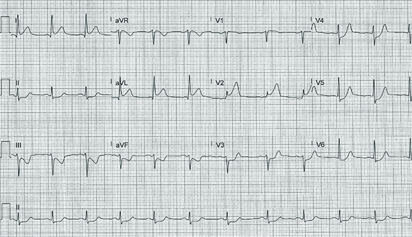
A 61-year-old male with past medical history of hypertension and hyperlipidemia presents to the emergency department with 8/10 substernal chest pain. What is your interpretation of his ECG?
Answer
This ECG shows normal sinus rhythm at 68 bpm, left axis deviation, normal intervals, STE in leads I, aVL, and V1-V2, and STD in leads II, III, aVF, and V3-V6.
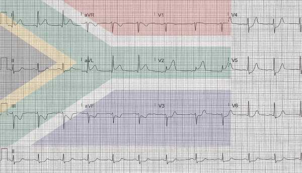
The pattern of STE and STD in this ECG is consistent with a high lateral MI. This pattern is sometimes called the South African Flag sign because the leads that show STE (leads I, aVL, and V2) and STD (lead III) fit into the central green coloration of the South African flag (see Figure 1).
Figure 1. The South African Flag sign: the green coloration of the South African flag overlays the leads that show STE (leads I, aVL, and V2) and STD (lead III) seen in a high lateral MI
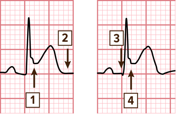
STEMI is defined as new, or presumed new, STE in ≥ 2 anatomically contiguous leads in patients with suspected ACS. The ST-segment should be measured at the J-point and compared to the isoelectric baseline.
In patients with a stable baseline, the TP segment should be used.1 Otherwise, the onset of the QRS complex should be used1 (see Figures 2 and 3). Note that the presence of reciprocal STD supports, but is not required for, the diagnosis of STEMI.
The high lateral MI is an established ischemic pattern with the culprit lesion typically in the first diagonal branch of the LAD.2,3 The ECG seen with a high lateral MI can often show subtle STE in lead I that does not meet the AHA/ACC STEMI criteria, so it is important to recognize that STE in leads aVL and V2 with STD in lead III (and often aVF) is consistent with an occlusion of the 1st diagonal artery even though it does not meet traditional STEMI criteria since lead V2 is not considered contiguous with lead aVL.4,5
Conclusion
This patient underwent emergent coronary angiography, which showed a 100% occlusion of the 1st diagonal artery and a 90% occlusion of the mid LAD; both occlusions were successfully treated with stent placement.
STEMI Learning Points
- STEMI is defined by new, or presumed new, STE at the J point in ≥ 2 anatomically contiguous leads in the absence of LVH by voltage pattern, LBBB, or ventricular paced rhythm
- ≥ 2.5 mm in men < 40 years old and ≥ 2 mm in men ≥ 40 years old in leads V2-V3 (or an increase of ≥ 1 mm when compared to baseline ECG)
- ≥ 1.5 mm in women in leads V2-V3 (or an increase of ≥ 1 mm when compared to baseline ECG)
- ≥ 1 mm in all other leads
- ST-segment is measured from the isoelectric baseline, typically the TP segment
- Reciprocal STD are not required for the diagnosis of STEMI, but their presence does increase the likelihood of the diagnosis of STEMI
- New LBBB is no longer considered a STEMI equivalent
- Significant STE that does not meet the traditional distribution (ie, ≥ 2 anatomically contiguous leads) or STE that does not meet amplitude criteria in a presentation concerning for ACS may still represent coronary artery occlusion and warrant consideration for emergent coronary reperfusion
References
- Thygesen K, Alpert JS, Jaffe AS, et al. Fourth Universal Definition of Myocardial Infarction. Circulation. 2018;138:e618-e651.
- Durant E, Singh A. Acute first diagonal artery occlusion: a characteristic pattern of ST elevation in noncontiguous leads. Am J Emerg Med. 2015;33(9):1326.
- Sclarovsky S, Birnbaum Y, Solodky A, Zafrir N, Wurzel M, Rechavia E. Isolated mid-anterior myocardial infarction: a special electrocardiographic sub-type of acute myocardial infarction consisting of ST-elevation in non-consecutive leads and two different morphologic types of ST-depression. Int J Cardiol. 1994;46(1):37-47.
- Monahan M, Sreenivasan S. A Red Flag. Circulation. 2020;142(19): 1871-1874.
- littmann L. South African flag sign: a teaching tool for easier ECG recognition of high lateral infarct. Am J Emerg Med. 2016;34(1):107-109.