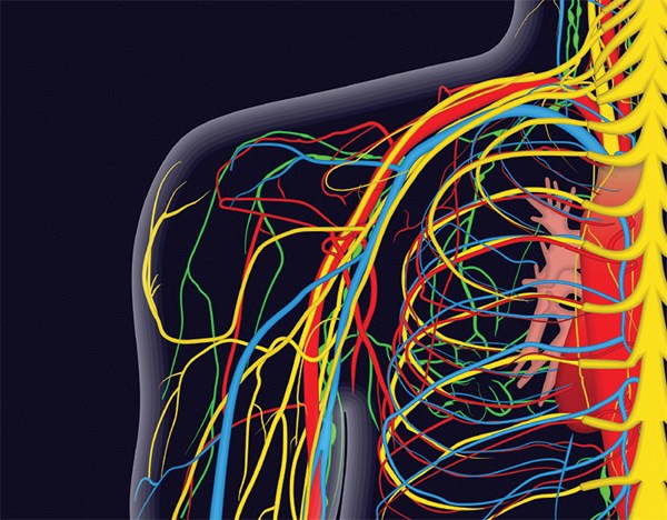
Interscalene nerve blocks have recently been popularized as a technique to facilitate shoulder reductions in the emergency department.
The procedure involves recognizing the brachial 5th, 6th, and 7th roots of the brachial plexus where they lie between the anterior and middle scalene muscles on the inferior/anterior neck. After the 5-7th roots are visualized, anesthetic is infiltrated around the nerves. This procedure has been shown to be of particular utility in shoulder reductions in the ED, with multiple studies illustrating decreased emergency room length of stay with the use of regional anesthesia compared to procedural sedation.1 While this is an important finding, especially among emergency physicians working in overcrowded EDs with long wait times, it is important that we consider the risks of this procedure and appropriate patient selection prior to popularizing its use, just as we have done with procedural sedation. While the benefits of the procedure are being promoted, the risks of the procedure have not been fully explored in the emergency medicine literature.
Interscalene nerve blocks were developed for analgesia prior to shoulder surgeries. As emergency physicians have broadened their skill sets with ultrasound (US)-guided regional nerve blocks, the procedure has become a tool available in the ED. When performed under direct real time visualization, there has been an increase in its success rate in the hands of less experienced physicians, and there has been a significant decrease in the potential risks of the procedure.2
Traditionally, interscalene blocks are performed under US guidance in the emergency department. This should only be considered after complete neuromuscular examination of the extremity, as regional anesthesia will limit any further assessment after administration. A complete view of the brachial plexus should be obtained in the interscalene position with administration of anesthetic performed under in-line visualization until envelopment of the brachial plexus is identified. Choice of anesthetic agent varies given the different length of actions; however, use of shorter-acting anesthetics seems to make sense in the setting of a short procedure with limited post-procedural pain. Prior to anesthetic administration, it is important to identify all important nearby structures to ensure these are avoided during the procedure.
Vasculature
The addition of US guidance and Doppler imaging has greatly assisted in the identification of vasculature in the area. Prior to administration of anesthetic, it is important to identify the carotid artery and the internal jugular vein. These are often out of the in-line view as they are more medial structures, but should be identified prior to initiation so that the performing physician can be aware and comfortable with their location. These should be avoided throughout the procedure, not only to decrease risk of bleeding or vessel injury, but also to ensure against inadvertent intravascular injections of anesthetic which can have a dose-dependent direct cardiovascular toxic effect.
Phrenic Nerve
While the phrenic nerve is often a considered structure in this area, its vicinity cannot be appreciated enough, especially when considering potential candidates for this procedure. As the phrenic nerve courses along the brachial plexus on its path to innervating the diaphragm, consideration of its paralysis or injury must be emphasized. Paralysis of the phrenic nerve secondary to anesthetic administration or direct injury can lead to hemiparalysis to the diaphragm. This is thought to be more directly correlated with anesthetic volume and cranial spread of anesthesia along the muscle fascia rather than secondary to direct injury.3
Several studies have shown decreased pulmonary function in the majority of patients post-interscalene blocks, suggesting that phrenic nerve paralysis can be an expected consequence of this procedure.4 There was no observed difference in this rate in comparison of the anterior or posterior US-guided approach. It is postulated that decreasing volumes of anesthetic may decrease this effect.4
While this may not have a huge role in otherwise healthy individuals, it can have drastic effects on ventilation in patients with already limited pulmonary function. Its effect on obese patients, those with hypoventilation, and those with primary lung disease (especially with oxygen requirements) must be considered prior to the procedure.
Dorsal Scapular Nerve
Although phrenic nerve injury is a well-known and respected possible complication of interscalene blocks, the long thoracic and dorsal scapular nerves are less often discussed. The dorsal scapular nerve (DSN) supplies innervation to the levator scapulae and rhomboid muscles. It is derived from the 5th cervical nerve root and can be identified via US at the 6th cervical root level, usually as a hyperechoic structure within the middle scalene muscle.5
Injury can lead to a chronic pain syndrome in these patients, with upper back and shoulder pain and varying levels of functional impairment.6 Traditionally, injury to the scapular nerve was avoided by the use of muscle stimulation/twitching, which is not usually utilized in the emergency department. This makes awareness and US identification all the more important so this structure can be avoided.
Long Thoracic Nerve
The long thoracic nerve (LTN) innervates the serratus anterior and is derived from the 5th and 6th cervical roots. It runs in close proximity to the DSN as described above, but is generally deeper, usually between the 6th and 7th cervical roots within or close to the middle scalene muscle. Injury of LTN can also contribute to a chronic pain syndrome with serratus anterior palsy. This can impair shoulder elevation and can lead to impingement syndromes.6
It is important to consider all structures near the brachial plexus when performing interscalene blocks. One Korean study showed that during a standard US-guided posterior brachial plexus block approach, the DSN was encountered as much as 60% of the time, and the long thoracic nerve was encountered up to 21% of the time.7 While this finding was identified under nerve stimulation and may overestimate associated risk of injury, it is important to consider this as a very viable complication of the procedure, even under US guidance, especially when the nerves are not identified prior to performing the procedure.
The utility of interscalene blocks in shoulder reductions is being recognized in the emergency department. It is important to be clear of surrounding neurovascular structures and to understand potential complications of this procedure prior to adopting it. Vascular structures should always be identified and avoided under US guidance. Potential patients should be screened for underlying pulmonary disease prior to the administration of anesthetic as phrenic nerve paralysis can be an expected complication of the procedure. Being able to recognize the DSN and LTN can help avoid damage to these important structures.
References
1. Blaivas M, Adhikari S, Lander L. A Prospective Comparison of Procedural Sedation and Ultrasound-guided Interscalene Nerve Block for Shoulder Reduction in the Emergency Department. Acad Emerg Med. 2011;18(9):922-927.
2. Raeyat Doost E, Heiran MM, Movahedi M, Mirafzal A. Ultrasound-guided interscalene nerve block vs procedural sedation by propofol and fentanyl for anterior shoulder dislocations. Am J Emerg Med. 2017;35(10):1435-1439.
3. Neal JM, Gerancher JC, Hebl JR, Ilfeld BM, McCartney CJ, Franco CD, Hogan QH. Upper extremity regional anesthesia: essentials of our current understanding, 2008. Reg Anesth Pain Med. 2009;34(2):134-170.
4. Bergmann L, Martini S, Kesselmeier M, et al. Phrenic nerve block caused by interscalene brachial plexus block: breathing effects of different sites of injection. BMC Anesthesiol. 2016;16(1):45.
5. Kim H. Dorsal scapular and long thoracic nerves during ultrasound-guided interscalene brachial plexus block. Asian J Anesthesiol. 2017;55(1):26-27.
6. Saporito A. Dorsal scapular nerve injury: a complication of ultrasound-guided interscalene block. Br J Anaesth. 2013;111(5):840-841.
7. Kim YD, Yu JY, Shim J, Heo HJ, Kim H. Risk of Encountering Dorsal Scapular and Long Thoracic Nerves during Ultrasound-guided Interscalene Brachial Plexus Block with Nerve Stimulator. Korean J Pain. 2016 Jul;29(3):179-184.


