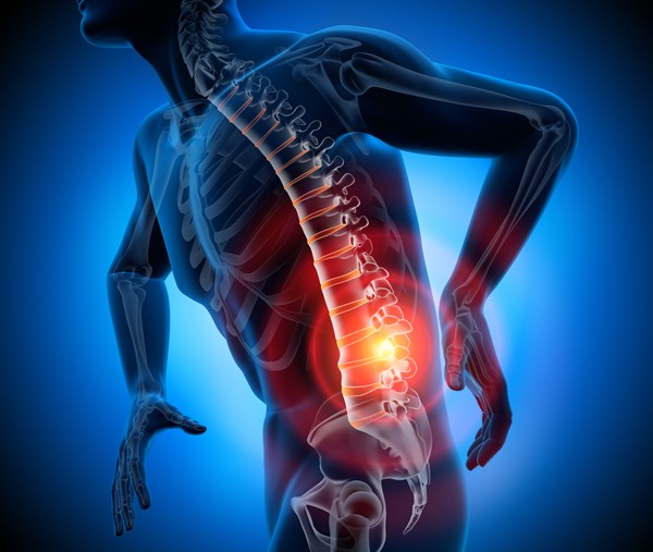
This case study presents a 75-year-old male who developed sudden quadriplegia while exercising. Initial stroke imaging was negative, and MRI revealed severe cervical spinal stenosis with spinal cord compression. The patient underwent emergent anterior cervical discectomy and fusion (ACDF) and was discharged to rehabilitation with residual deficits. This case highlights the importance of considering spinal cord injury (SCI) in the differential diagnosis of acute paralysis and the need for timely imaging and surgical management.
In this case study, we will discuss the epidemiology, risk factors, pathophysiology, classification, emergency management, and rehabilitation considerations of acute spinal cord injury, particularly non-traumatic causes such as degenerative cervical stenosis.
Patient History and Presentation
A 75-year-old male with a past medical history of hypertension, hyperlipidemia, chronic neck pain, and past alcohol abuse presented after a syncopal episode at a gym while performing “planks.” Emergency medical services found him supine on the floor, alert and oriented. He denied any trauma or fall and had no signs of head injury.
Upon arrival at the emergency department, vital signs were within normal limits: heart rate 61 bpm, respiratory rate 18/min, blood pressure 123/79 mmHg, oxygen saturation 98% on room air, and temperature 36.4°C. Neurologically, he exhibited no facial droop, aphasia, or dysarthria. However, he had sudden quadriplegia with intact sensation in all extremities and no pathological reflexes. A stroke alert was triggered due to a National Institutes of Health Stroke Scale (NIH SS) score of 19.
Initial imaging, including non-contrast CT of the head, CT angiography of the head and neck, and CT perfusion studies, revealed no acute ischemic or hemorrhagic stroke and no large vessel occlusion. An emergent cervical spine MRI revealed severe spinal canal stenosis at C3–C4 with significant disc herniation and spinal cord compression, along with additional multilevel stenosis from C4–C6. There was also significant soft tissue edema consistent with anterior longitudinal ligament injury.
The patient underwent emergent neurosurgical intervention with a C3–C6 anterior cervical discectomy and fusion (ACDF). He was managed postoperatively in the intensive care unit for seven days and subsequently discharged to a rehabilitation facility on postoperative day ten. At discharge, significant motor deficits persisted in all four limbs, with complete motor paralysis in the lower extremities and severe weakness in the upper extremities.
Discussion
Spinal cord injury (SCI) is a potentially life-altering neurological emergency associated with high rates of morbidity and long-term disability. Globally, the incidence of SCI ranges from 10 to 80 cases per million people annually, with a predominance in high-income countries due to higher rates of motor vehicle accidents and trauma. In the United States, roughly 17,000 new cases occur each year, and males constitute about 78% of all cases. Notably, in elderly populations, non-traumatic etiologies such as cervical spondylosis and spinal stenosis have become more prominent causes of SCI.1
This patient’s presentation aligns with a non-traumatic cervical spinal cord injury, which can be more subtle and often misdiagnosed initially. Risk factors for non-traumatic SCI include older age, male sex, a history of chronic neck pain, cervical spondylosis, degenerative joint disease, and osteoporosis. Physical activity involving hyperextension of the neck, as seen in this case during “planks,” may precipitate cord compression in individuals with predisposing spinal canal narrowing. Plank exercises, although generally safe, require sustained isometric contraction and spinal alignment; in elderly individuals with cervical stenosis, the posture can promote neck extension and increased axial load, triggering anterior disc herniation or ligamentous strain. Such biomechanical stress can acutely narrow the spinal canal and lead to cord impingement, particularly in patients with preexisting cervical degeneration. This mechanism has been described in rare cases of cervical spinal injury during low-impact activities like yoga or core training, emphasizing the vulnerability of the aging spine to mechanical insults even without trauma.2
Pathophysiologically, SCI occurs in two main phases. The primary injury is caused by direct mechanical disruption of axons, contusion, or compression due to fractures, herniated discs, or ligamentous injuries. This initial event is followed by a secondary phase involving ischemia, inflammatory mediator release, excitotoxicity, oxidative stress, and apoptosis.3,4,5 These secondary events expand the area of damage and increase functional impairment over time. Prompt surgical decompression and maintenance of adequate spinal cord perfusion are critical in limiting the extent of secondary injury.6
Spinal cord injuries can be classified as complete or incomplete. In complete SCI, there is total loss of motor and sensory function below the level of injury, including the absence of sacral sparing, and recovery is typically poor. In incomplete SCI, some motor or sensory function remains, particularly in the sacral segments, and the prognosis is comparatively more favorable. Several specific syndromes are described under incomplete SCI. Anterior cord syndrome, usually due to infarction or anterior spinal artery compromise, results in bilateral motor paralysis and loss of pain and temperature sensation, with preservation of proprioception and vibration sense. Central cord syndrome, often associated with hyperextension injuries in elderly patients with preexisting cervical spondylosis, presents with greater weakness in the upper extremities than in the lower extremities.1
The patient’s presentation of sudden quadriplegia with intact sensation and absence of trauma suggests an incomplete cervical SCI. The findings on MRI, including severe canal stenosis at multiple cervical levels and significant spinal cord compression with soft tissue swelling, are consistent with central cord syndrome. However, the bilateral and symmetric weakness also suggests a more diffuse cervical myelopathy rather than a pure syndrome.
Management
The management of acute spinal cord injury (SCI) begins in the emergency department and continues through surgery, intensive care, and rehabilitation.7 The goals are to stabilize the spine, minimize secondary injury, maintain neurologic function, and optimize recovery. Acute cervical SCI, such as in this case, requires immediate attention to airway, breathing, and circulation, along with neuroprotection and surgical planning.
One of the first and most important interventions is hemodynamic support. Patients with SCI, particularly at or above the T6 level, are at risk for neurogenic shock, characterized by hypotension and bradycardia due to disruption of sympathetic pathways. Guidelines recommend maintaining a mean arterial pressure (MAP) of 85–90 mmHg for at least 5 to 7 days post-injury to improve spinal cord perfusion and support neurological recovery.7,8
Respiratory complications are among the leading causes of early mortality in cervical SCI. Injuries above the C5 level may impair diaphragmatic function, requiring mechanical ventilation or tracheostomy. Pulmonary complications such as atelectasis, pneumonia, and respiratory failure are common and require aggressive management.1
Cardiovascular monitoring is equally critical. Patients can experience profound bradycardia, even asystole, due to unopposed vagal tone. In some cases, atropine or temporary pacing may be necessary.8 Autonomic instability also increases the risk for arrhythmias and orthostatic hypotension, especially during mobilization.
Neuroimaging, particularly MRI, is the gold standard for diagnosis in patients with unexplained neurological deficits when CT findings are negative.6 This patient’s MRI, showing multilevel cervical stenosis with cord compression and ligament injury, prompted emergent surgical decompression, which is consistent with guidelines.
Surgical intervention—most effectively within 24 hours—has been associated with improved neurological outcomes, particularly in incomplete SCI.1,6 The patient underwent prompt anterior cervical discectomy and fusion (ACDF) at levels C3–C6, consistent with evidence-based best practices for acute cord compression. The goal of surgery is to decompress the cord, stabilize the spine, and prevent further injury.
The use of high-dose corticosteroids, specifically methylprednisolone, remains controversial. It is not routinely recommended due to increased risks of gastrointestinal bleeding, infections, and delayed wound healing. However, it may be considered within 8 hours of traumatic SCI in select cases, but not in atraumatic causes like degenerative spinal stenosis.8
In the ICU post-operatively, prevention of secondary complications is paramount. Patients should receive venous thromboembolism (VTE) prophylaxis with low molecular weight heparin within 72 hours unless contraindicated.8 Additionally, stress ulcer prophylaxis with proton pump inhibitors or H2 blockers is warranted, particularly in patients on steroids or mechanical ventilation.
Bladder and bowel management is initiated early with an indwelling catheter and transitioned to intermittent catheterization. Bowel programs are introduced to maintain regular elimination and prevent impaction. Pressure injury prevention involves the use of specialized mattresses and routine repositioning to prevent skin breakdown.8
Prognosis
Rehabilitation begins as early as possible. Once stabilized, patients should be transferred to a comprehensive SCI rehabilitation unit. Recovery is most promising in incomplete injuries. Rehabilitation focuses on mobility, strength, activities of daily living (ADLs), spasticity control, and psychological support.9
Prognosis in SCI depends on several factors, including the severity and level of injury, time to surgical intervention, age, comorbidities, and access to rehabilitation.6,9 Patients with incomplete injuries such as central cord syndrome often have good prognosis for functional improvement, particularly in lower extremities, but may continue to experience persistent upper extremity weakness. In this case, residual quadriparesis at discharge suggests significant cervical cord involvement, but ongoing rehabilitation will likely result in some neurological recovery.
Conclusion
In conclusion, this case illustrates how spinal cord injury, particularly from non-traumatic degenerative etiologies, can mimic acute neurological emergencies like stroke. Prompt recognition, neuroimaging, and neurosurgical intervention are crucial to improve functional outcomes. Emergency physicians must maintain a high index of suspicion for spinal pathology in older adults presenting with acute paralysis, especially in the absence of cranial neurological findings.
References
- Ahuja CS, Wilson JR, Nori S, et al. Traumatic spinal cord injury. Nat Rev Dis Primers. 2017;3:17018.
- Dalbayrak S, Yaman O, Yilmaz M, et al. Acute central cervical cord syndrome in a yoga practitioner: Case report and review of the literature. Turk Neurosurg. 2015;25(5):818-821. doi:10.5137/1019-5149.JTN.12346-14.1
- Anjum A, Yazid MD, Fauzi Daud M, et al. Spinal Cord Injury: Pathophysiology, Multimolecular Interactions, and Underlying Recovery Mechanisms. Int J Mol Sci. 2020;21(20):7533.
- Quadri SA, Farooqui M, Ikram A, et al. Recent update on basic mechanisms of spinal cord injury. Neurosurg Rev. 2020;43(2):425-441.
- Karsy M, Hawryluk G. Modern Medical Management of Spinal Cord Injury. Curr Neurol Neurosci Rep. 2019;19(9):65.
- Chay W, Kirshblum S. Predicting Outcomes After Spinal Cord Injury. Phys Med Rehabil Clin N Am.2020;31(3):331-343.
- Picetti E, Marchesini N, Biffl WL, et al. The acute phase management of traumatic spinal cord injury (tSCI) with polytrauma: A narrative review. Brain Spine. 2024;4:104146.
- UpToDate. Acute traumatic spinal cord injury.
- Sharif S, Ali MYJ. Outcome Prediction in Spinal Cord Injury: Myth or Reality. World Neurosurg. 2020;140:574-590.


