


POCUS is a key diagnostic tool that can allow emergency physicians to diagnose a range of injuries. This case highlights the utility of bedside point-of-care ultrasound in diagnosing a ruptured Achilles tendon.
This paper was edited by:
Madeleine Silverstein, MD
Department of Emergency Medicine
UT Southwestern Medical Center
Vietvuong Vo, MD
Assistant Professor, Department of Emergency Medicine
UT Southwestern Medical Center
Ayomide Isola-Gbenla
Texas A&M School of Medicine
Class of 2025
Sean Beckman, DO
Department of Emergency Medicine
Rutgers New Jersey Medical School
HISTORY OF PRESENT ILLNESS
A 35-year-old male with no significant past medical history presented to the emergency department (ED) with left calf pain that started while playing basketball that evening. The patient described feeling a sudden "pop" in his distal left calf, immediately followed by sharp pain and inability to bear weight. He denied any other injuries, falls, loss of consciousness, or head trauma.
PHYSICAL EXAMINATION
His vital signs were a blood pressure of 118/66 mmHg, heart rate of 111 bpm, temperature of 99.2°F, respiratory rate of 18 breaths/minute, and SpO2 96% on room air. He appeared in no acute distress and had no obvious external signs of traumatic injury. His exam was notable for tenderness to palpation at the distal posterior left gastrocnemius along the Achilles tendon. He was unable to actively dorsiflex his foot, however sensation was intact throughout the lower extremity. Dorsalis pedis and posterior tibial pulses were palpable and 2+ bilaterally. The Thompson's test was positive, with no plantarflexion observed upon squeezing the left calf while lying prone, suggesting an acute Achilles tendon rupture.
LABS AND IMAGING
X-rays were performed of the left foot and ankle (Image 1). Both were negative for acute fracture, dislocation, or foreign body.
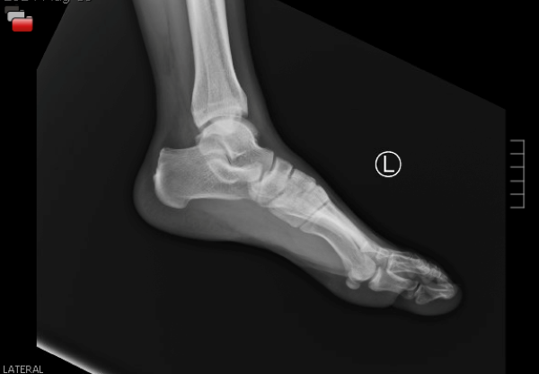 Image 1. Left ankle x-ray
POCUS
Point-of-care ultrasound (POCUS) was performed to evaluate the integrity of the Achilles Tendon. Using the linear probe in transverse and sagittal orientations, a disruption of the tendon was visualized with a surrounding anechoic fluid collection consistent with a hematoma, indicative of acute tendon tear. (Image 2,3. Video 1,2)
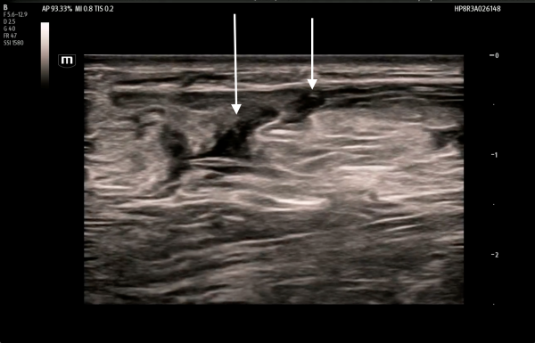
Image 2.Sagittal view - disruption of the tendon with a surrounding fluid collection
POCUS FTW Achilles Tendon - Video 1.mp4
Video 1. Sagittal Clip - disruption of the tendon with a surrounding fluid collection
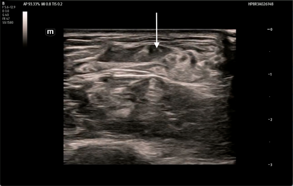
Image 3. Transverse view - disruption of the tendon with a surrounding fluid collection
POCUS FTW Achilles Tendon - Video 2.mp4
Video 2. Transverse Clip - disruption of the tendon with a surrounding fluid collection
DISCUSSION
The Achilles tendon is the most commonly ruptured tendon, with 40 cases per 100,000 annually.1 This case highlights the utility of bedside point-of-care ultrasound in the emergency department for diagnosing musculoskeletal injuries. Historically, Thompson’s test alone has been used to diagnose Achilles tendon injuries in the ED. It has an estimated sensitivity of 96% and a specificity of 93%.2 Despite these metrics, Achilles tendon ruptures are misdiagnosed as ankle sprains in up to 20–25% of cases.3 Misdiagnosis of these injuries may result in poor functional outcomes and increased risk of re-rupture, as untreated patients continue full ankle motion, exacerbating injury severity.4 Proper diagnosis of Achilles tendon rupture significantly impacts patient management compared to the common misdiagnosis of ankle sprain. Management options include both operative and nonoperative approaches.5 Conventional non-operative care involves immobilization in a plantarflexed splint or 3D boot.5
We propose that implementing both bedside clinical exams, such as Thompson’s test, and POCUS can significantly reduce the misdiagnosis of Achilles tendon ruptures. Ultrasound has been shown to have high sensitivity (94.8%) and specificity (98.7%) for diagnosing complete Achilles tendon ruptures, providing both patient and provider with a confirmed diagnosis in the ED.4 On ultrasound, a normal Achilles tendon should appear organized with the parallel, tightly packed fibers. Injury to the tendon disrupts this organization, manifesting as loosening of the fibers, or appearing like the end of a frayed rope. Edema and bleeding appear as heterogeneous, hypoechoic changes or anechoic fluid pockets. The disruption and edema may both contribute to abnormal tendon thickening greater than 1cm in diameter. Partial ruptures may have some or all of these changes but with some fibers still intact. Complete ruptures demonstrate full loss of normal tendon structure with a visible gap, while partial ruptures typically exhibit areas of disrupted fibers interspersed with intact fibers and focal hypoechoic changes. The unaffected side should be used for comparison.4 Although MRI is considered the gold standard for diagnosing tendon injuries and is important for operative planning, it is costly, time-consuming, and less accessible in emergency settings. Ultrasound serves as a practical and effective diagnostic tool in the emergency department, eliminating barriers to treatment and evaluation.
HOSPITAL COURSE and CASE RESOLUTION
The patient was immobilized in a plantarflexed short leg splint. Orthopedic surgery was consulted and recommended outpatient follow-up. Subsequent Orthopedic evaluation led to surgical Achilles tendon repair. The patient has been doing well post-operatively.
REFERENCES
- Lemme NJ, Li NY, DeFroda SF, Kleiner J, Owens BD. Epidemiology of Achilles Tendon Ruptures in the United States: Athletic and Nonathletic Injuries From 2012 to 2016. Orthop J Sports Med. 2018 Nov 26;6(11):2325967118808238. doi: 10.1177/2325967118808238. PMID: 30505872; PMCID: PMC6259075.
- Schwieterman B, Haas D, Columber K, Knupp D, Cook C. Diagnostic accuracy of physical examination tests of the ankle/foot complex: a systematic review. Int J Sports Phys Ther. 2013 Aug;8(4):416-26. PMID: 24175128; PMCID: PMC3812842.
- Shamrock AG, Dreyer MA, Varacallo MA. Achilles Tendon Rupture. [Updated 2023 Aug 17]. In: StatPearls [Internet]. Treasure Island (FL): StatPearls Publishing; 2025 Jan-. Available from: https://www.ncbi.nlm.nih.gov/books/NBK430844/#
- Amir Aminlari, Jennifer Stone, Ryan McKee, Rachna Subramony, Adam Nadolski, Vaishal Tolia, Stephen R. Hayden. Diagnosing Achilles Tendon Rupture with Ultrasound in Patients Treated Surgically: A Systematic Review and Meta-Analysis. The Journal of Emergency Medicine. Volume 61, Issue 5, 2021, Pages 558-567,ISSN 0736-4679, https://doi.org/10.1016/j.jemermed.2021.09.008.
- Park SH, Lee HS, Young KW, Seo SG. Treatment of Acute Achilles Tendon Rupture. Clin Orthop Surg. 2020 Mar;12(1):1-8. https://doi.org/10.4055/cios.2020.12.1.1
- Atta M, Jafari S, Moore K. Analyzing the use of ultrasound: achilles tendon rupture. Open J Emerg Med. 2019;7:41–7. https://doi.org/10.4236/ojem.2019.73005.