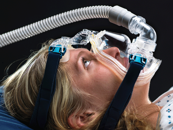
It is important for the emergency physician to be comfortable directing and troubleshooting NIPPV in order to promote optimal patient outcomes.
Paramedics are called to the home of a 63-year-old female with shortness of breath. They find an older woman who is alert but appears anxious, and is in moderate respiratory distress. She is unable to effectively give a detailed medical history due to dyspnea. Two unmarked metered dose inhalers are noted at the patient's bedside. The patient is started on non-invasive positive pressure ventilation (NIPPV) in bi-level positive airway pressure (BiPAP) mode with in-line nebulized albuterol/ipratropium. Upon arrival to the emergency department (ED), the EMS crew report that the patient is much improved. Should you continue NIPPV? If so, what NIPPV parameters should you specify?
Introduction
NIPPV in the emergency department is a common respiratory intervention modality that is increasingly initiated in the prehospital arena. It has been shown to be an effective means of improving multiple etiologies of dyspnea while potentially avoiding the morbidities associated with endotracheal intubation. Because of its frequent utility, it is important for the emergency physician to be comfortable directing and troubleshooting NIPPV in order to promote optimal patient outcome. Herein, we will review common modes of NIPPV, pathophysiology, etiology-specific considerations, and troubleshooting.
Definitions
NIPPV is a broad term used to describe several methods of ventilation management. It often refers to positive pressure delivered by air-tight facial or nasal mask. Two common modes are continuous positive airway pressure (CPAP) and BiPAP.1 CPAP provides a constant positive pressure on inspiration and expiration. BiPAP provides a baseline pressure, as well as a second, elevated inspiratory pressure triggered by the patient's inspiratory effort. The lower pressure delivered on expiration is referred to as expiratory positive airway pressure (EPAP) and is analogous to positive end-expiratory pressure (PEEP) delivered by a traditional ventilator. The higher pressure delivered with inspiration is termed inspiratory positive airway pressure (IPAP). The difference between these two values is the pressure support (PS). A minimum respiratory rate can be set on BiPAP, whereas no rate can be set on CPAP. The FiO2 can be adjusted in both modes. Because this is a patient-driven modality, the respiratory rate and the tidal volume delivered are determined by the patient's effort, while IPAP and EPAP are controlled by the ventilator.2
Pathophysiology
There are several beneficial changes in pathophysiology and lung mechanics that may occur with the use of NIPPV. These vary depending on the underlying disease, the severity of the physiologic derangement, and the mode of NIPPV.
In hypoxemic respiratory failure, the extrinsic PEEP delivered by NIPPV can improve alveolar recruitment and gas exchange. Specifically, NIPPV can deliver optimal FiO2, recruit collapsed alveoli, improve V/Q mismatch and allow improved flow of fluid from alveoli back into circulation.3
In the setting of acute exacerbation of congestive heart failure (AECHF) with acute cardiogenic pulmonary edema (ACPE), cardiac output often improves due to changes in preload and afterload. The additional PEEP delivered by NIPPV increases intrathoracic pressure causing a decrease in venous return, which prevents overfilling of the right heart. Increased intrathoracic pressure decreases left ventricular transmural pressure, thus decreasing afterload.4 The net effect of these changes results in decreased stretching of cardiac muscle fibers, allowing them to function at a more favorable portion of the Frank-Starling curve.
In the setting of hypercapnic respiratory failure associated with chronic obstructive pulmonary disease (COPD), the pressure support provided by BiPAP decreases work of breathing and improves ventilation.3 Further, the positive expiratory pressures provided by either mode can help prevent both air trapping and dynamic hyperinflation.
Indications
When utilized optimally, NIPPV improves oxygenation, work of breathing, respiratory mechanics, and cardiac function. It should be considered for any dyspneic patient presenting to the ED assuming no contraindications exist (Figure 1).5-6 Among the most supported indications for NIPPV are ACPE and COPD.7
COPD
NIPPV is commonly used for patients with COPD to augment ventilation and decrease work of breathing in those with moderate-to-severe exacerbations, and is used in conjunction with standard beta-2-agonists and anticholinergic therapy.8 It is important for providers to pay careful attention to tidal volumes (these are patient-dependent and not set on the machine, but should be between 5-8cc/kg ideal body weight) as well as unintentional mask leak. In patients presenting with an uncompensated respiratory acidosis (and elevated PCO2), the IPAP should be raised to increase the tidal volumes and improve minute-ventilation. The patient may not tolerate an IPAP above 10 cm H2O initially, but usually with adequate bedside coaching the IPAP can be steadily increased.
AECHF
NIPPV is a beneficial adjunct to nitrates and typical medical therapy in AECHF with ACPE. As discussed above, the increased intrathoracic pressures from NIPPV can improve cardiac function in the diseased heart. While both modes of NIPPV demonstrate comparable benefits, CPAP is the modality with the most consistently reproduced benefits in the setting of ACPE.5,9,10
NIPPV Setup
All emergency medicine providers should be comfortable with the set-up and management of NIPPV. For example, it may be necessary to quickly transition a patient from pre-hospital NIPPV when respiratory therapy is not immediately available. ED respiratory therapists are a great resource for learning where the equipment is located and how a particular NIPPV device is managed. A critical and seldom discussed aspect of setup is coaching. Placing a mask on a patient already struggling to breathe can induce panic and subsequent failure of NIPPV. In the NIPPV naive patient, you may be “teaching someone to swim as they are drowning,” so to speak. Going through the process at the patient's bedside and explaining each step while encouraging and calming them will allow the patient the best chance at NIPPV success. For patients unfamiliar with NIPPV, begin with low initial pressures and then titrate settings according to the patient's pathology and response to therapy (Figure 2).
In patients who arrive on pre-hospital NIPPV, it is important to assess the duration of therapy and patient's response prior to arrival. When obtaining initial history, consider the differing nomenclature between traditional ventilators and NIPPV machines. In prehospital jargon,“10 and 5” usually means: 15 cm H2O IPAP, 5 cm H2O EPAP, and PS of 10 cm H2O. When referring to a BiPAP machine, this language actually means: 10 cm H2O IPAP, 5 cm H2O of EPAP, and PS of 5 cm H2O. By accurately obtaining this history, the emergency physician is then prepared to best tailor settings.
Troubleshooting
Because there is no “one-size-fits-all” approach to NIPPV use, providers must quickly establish working differential diagnoses and adjust changes in ventilator settings according to the most likely diagnosis. For instance, if the patient likely has CHF with ACPE, choose settings that most benefit oxygenation. To do this, maximize extrinsic PEEP by increasing CPAP or EPAP values, while maintaining PS relatively low. If your patient requires augmented ventilation, as would likely be the case with COPD, choose settings that maximize PS by increasing IPAP. Frequent coaching, monitoring and titration of settings will likely be required until clear signs of improvement are noted.
Sometimes adequate tidal volumes may not be achieved due to unintentional mask leak. Assuring correct mask size and adjustment is necessary to optimize patient comfort and to minimize air leak around the mask. The mask fit should be tight enough to form a seal between the facial skin and the mask interface, but should be loose enough to be able to pull away and reposition if needed. A patient with excessive facial hair or facial asymmetry will be much more difficult to adequately seal and deliver optimal pressure. Make sure the patient is as comfortable as possible and offer frequent reassurance.
NIPPV Failure
Key clinical markers to re-evaluate frequently on NIPPV patients include: mental status, respiratory rate/work of breathing, vital signs, tidal volumes, and ABG/VBG values.11 If the patient is not improving clinically with frequent re-evaluation and optimal NIPPV management, the provider should not hesitate to intubate if indicated. In the setting that optimal ventilatory parameters are delivered, there should be significant improvement in the first hour of delivery. However, each patient should be considered individually as the timing, etiology, and severity of the respiratory distress can contribute to NIPPV failure or success.12
If the patient is not improving with levels of applied PEEP (>10-12 cm H2O) the patient may not have a condition amenable to NIPPV and intubation is likely necessary (Figure 3).7 It is possible that no recruitable alveoli remain, and increasing extrinsic pressure may serve only to over-distend functioning alveoli leading to harmful inflammation. Importantly, discontinue NIPPV in any patient with signs that they may be developing a preload dependent state, as this can decrease venous return and hinder cardiac output.
Figure 1. Contraindications to NIPPV
1. Inability to protect airway
2. Impaired consciousness or inability to cooperate
3. Respiratory or cardiac arrest
4. Hemodynamic instability
5. Serious facial injury or deformity
6. Pneumothorax
7. Upper Airway obstruction
8. Multi-organ failure
9. Recent esophageal anastomosis
© 2011-2017, American College of Emergency Physicians, Reprinted with Permission.
Conclusion
As the initiation of pre-hospital NIPPV increases, the ability of the emergency physician to comfortably transition the patient for continued dynamic therapy is invaluable. In order for this process to be as seamless as possible, a thorough understanding of disease pathology, respiratory mechanics, NIPPV setup, and troubleshooting is paramount. Finally, NIPPV is not a substitute for establishing a definitive airway when indicated. Do not hesitate to intubate when necessary.
References
- Moy HP, Bruton B. Evidence-Based EMS: Out-of-Hospital BiPAP vs. CPAP. EMS World. http://www.emsworld.com/article/12145134/evidence-based-ems-out-of-hospital-bipap-vs-cpap. Published December 31, 2015. Accessed November 13, 2016.
- British Thoracic Society Standards of Care Committee. Non-invasive ventilation in acute respiratory failure. Thorax. 2002;57(3):192-211.
- Duke GJ, Bersten AD. Non-invasive ventilation for adult acute respiratory failure. Part I. Crit Care Resusc. 1999;1(2):187-198.
- Wright BJ, Slesinger TL. Noninvasive Positive Pressure Ventilation. In: Farcy DA, Chiu WC, Marshall JP, Osborn TM. eds. Critical Care Emergency Medicine, 2nd ed. New York, NY: McGraw-Hill; 2016.
- Li H, Hu C, Xia J, Li X, Wei H, Zeng X, et al. A comparison of bilevel and continuous positive airway pressure noninvasive ventilation in acute cardiogenic pulmonary edema. Am J Emerg Med. 2013;31(9):1322-1327.
- Burns B. Noninvasive ventilation a practical guide. Emerg Med. 2015;47(1):20-29.
- Ahn J, Pillow T. Focus On: Noninvasive Positive Pressure Ventilation In the Emergency Department. ACEP News. March 2010. https://www.acep.org/Clinical---Practice-Management/Focus-On--Noninvasive-Positive-Pressure-Ventilation-In-the-Emergency-Department. Accessed November 10, 2016.
- Ward, NS. Dushay,KM. Clinical concise review: mechanical ventilation of patients with chronic obstructive pulmonary disease. Crit Care Med. 2008;36:1614-1619.
- Peter JV, Moran JL, Phillips-Hughes J, Graham P, Bersten AD. Effect of non-invasive positive pressure ventilation (NIPPV) on mortality in patients with acute cardiogenic pulmonary oedema: a meta-analysis. The Lancet. 2006;367(9517):1155-1163.
- Collins SP, Mielniczuk LM, Whittingham HA, Boseley ME, Schramm DR, Storrow AB. The use of noninvasive ventilation in emergency department patients with acute cardiogenic pulmonary edema: a systematic review. Ann Emerg Med. 2006;48(3):260-269.
- Slesinger TL, Wright BJ. Chapter 6. Noninvasive Ventilation. In: Farcy DA, Chiu WC, Flaxman A, Marshall JP. eds. Critical Care Emergency Medicine. New York, NY: McGraw-Hill; 2012.
- Ozyilmaz E, Ugurlu AO, Nava S. Timing of noninvasive ventilation failure: causes, risk factors, and potential remedies. BMC Pulm Med. 2014;14(1):19.
- Carlson JN, Wang HE. Noninvasive Airway Management. In: Tintinalli JE, Stapczynski J, Ma O, Yealy DM, Meckler GD, Cline DM. eds. Tintinalli's Emergency Medicine: A Comprehensive Study Guide, 8e. New York, NY: McGraw-Hill; 2016.


