


A 7-year-old male with no past significant medical history presented to the emergency department, accompanied by his mother, for evaluation of bruising to the lower extremities that had been observed for the past week.
The mother reported she was initially not concerned about the bruising because the patient had started karate lessons. However, the patient reported to mom that he was not getting kicked in the legs.
A few days prior to presentation, the patient developed a large bruise to the right abdomen just above the hip. All of these bruises reportedly occurred without any trauma. This was when he was having a cough and rhinorrhea, which mom thought may have been allergies. The patient was reported to have an occasional nosebleed about once every other month, including one just in the previous week. Both the patient and mom denied fever, sore throat, myalgias, arthralgias, night sweats, weight loss, hemoptysis, hematuria, melena or hematochezia, bleeding gums, abdominal pain, nausea, vomiting, chest pain, shortness of breath, abnormal breath sounds, ear pain or discharge, foamy or frothy urine, changes in color of urine, changes in frequency of urination, rashes, or family history of bruising disorders. There was no recent antibiotic use or travel, and no recent known tick, mosquito, or other parasitic exposure. No new foods had been introduced to the patient’s diet. No new pets or chemical exposures were reported.
Nursing reported that petechiae appeared at the site of the blood pressure (BP) cuff after the BP was obtained.
The patient’s vital signs were stable. There were no labs drawn at the time of the initial evaluation.
A physical exam revealed a patient who appeared in no acute distress. He was attentive and interactive. Heart sounds were normal rate and regular without murmurs. Lungs were clear to auscultation with good chest expansion. The abdomen was soft and nontender. The spleen was not enlarged. There was no edema. The eyes had normal conjunctiva bilaterally. There were 6 small petechiae noted within the oropharynx. The patient had small areas of petechiae noted on bilateral anterior upper arms. Scattered petechiae were noted across the upper chest anteriorly. There was a large area of ecchymosis on the lower right side of the abdomen. He had innumerable ecchymotic lesions over the bilateral lower extremities, especially below the knees and anteriorly. There were no rashes on the palms or soles.
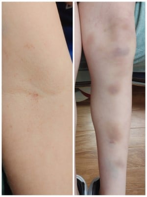
Bruising, lower extremities
Discussion
In this case, what would be your initial workup? The differential here should include malignancies such as myelodysplastic syndromes, leukemias, lymphomas; infections such as post-viral, HIV, Hepatitis C; drug-induced such as many antibiotics, seizure medications; and primary hematologic processes such as thrombotic thrombocytopenic purpura and disseminated intravascular coagulation. In this case, post-viral platelet disruption was thought to be the leading diagnosis.
Initial workup should include CBC, reticulocyte count, blood smear, fibrinogen, Von willebrand factor (VWF) activity, and a coagulation panel. In this case, the reticulocyte count was not ordered.
Our patient’s labs are shown (Table 1). A smear was not readily available but would typically be normal with variable platelet size, large platelets, and some giant platelets.
Table 1
|
WBC |
5.62 |
|
RBC |
4.03 L |
|
HGB |
11.5 |
|
HCT |
31.7 L |
|
MCV |
78.7 |
|
MCH |
28.5 |
|
MCHC |
36.3 H |
|
PLT |
1 LLL |
|
RDW |
12.3 |
|
Fibrinogen |
180 L |
|
PT |
11.1 |
|
INR |
1.0 |
|
COVID |
Not detected |
|
VWF activity |
Send out |
A diagnosis of virus-associated idiopathic thrombocytic purpura (ITP) was made.
The incidence of post-viral ITP is about 1 per 20,000 children a year. Two-thirds of children who develop ITP have had a recent viral illness. Occasionally, a virus such as EBV, varicella, or HIV (testing is not usually indicated) can be detected. The mechanism is not fully understood but typically results from increased platelet destruction and/or decreased production. A majority of children only develop minor bleeding, such as our patient with scattered ecchymosis, petechiae, and mucosal bleeding.
Treatment will usually be outpatient. The American Society of Hematology (ASH) suggests any patient with no or only minor bleeding be observed, plus/minus steroids or 2nd line medications, with hematology follow-up within 48 hours. This differs from the adult disposition. Any significant bleeding, of course, requires immediate inpatient treatment.
In patients with no or minor bleeding, ASH recommends observation over steroids or IVIG. If a decision is made to give steroids, ASH suggests a course of 2-4 mg/kg/day for 5 to 7 days. If there is a contraindication to steroids, then IVIG 1 g/kg x 1 can be given with the possibility of repeating dosing once. If there is severe, life-threatening bleeding, then IVIG with above dosing is indicated. In the event that symptoms persist for longer than 3 months, second-line agents — including thrombopoietin receptor agonists, rituximab, and/or splenectomy — may be indicated. All patients and parents should be given strict precautions to not participate in contact sports, collision activities, trampoline use, or any activity where there is higher risk for developing bleeding, especially intracranial bleeds. Children prone to falls and trauma, such as toddlers, may require treatment since they may not be able to follow these precautions.
Case Resolution
Our patient had non-severe bleeding and was discharged from the ED after a discussion with pediatric hematology. Guidance was given to the patient and mother to avoid activities that could lead to injury. Return precautions — including epistaxis, hematemesis, hemoptysis, hematochezia, and neurologic changes suggesting intracranial bleeding — were discussed. The patient followed up with hematology the next day, and a decision was made to administer a short course of oral steroids.
References
Kim TO, Despotovic JM. Pediatric immune thrombocytopenia (ITP) treatment. Annals of Blood. 2021;6:4-4. doi:10.21037/aob-20-96
Liebman HA. Viral-Associated Immune Thrombocytopenic Purpura. Hematology. 2008;2008(1):212-218. doi:10.1182/asheducation-2008.1.212
Neunert C, Vesely S, Kim T. Management of Immune Thrombocytopenia (ITP) ASH CLINICAL PRACTICE GUIDELINES IMMUNE THROMBOCYTOPENIA (ITP) POCKET GUIDE. https://www.hematology.org/-/media/Hematology/Files/Education/Clinicians/Guidelines-Quality/Documents/ASH-ITP-Pocket-Guide-FOR-WEB-1204.pdf
Rand, Ph.D ML, Fraser Wright, Ph.D J. Virus-associated Idiopathic Thrombocytopenic Purpura. Transfusion Science. 1998;19(3):253-259. doi:10.1016/s0955-3886(98)00039-3