


Intern year is full of teachable moments, as new physicians build repetitions seeing typical presentations of common chief complaints. But the true beauty of an intern year in emergency medicine comes when that intern finally develops a basic foundation of expediently recognizing and managing emergent life-threatening conditions, only to be swiftly confronted by a zebra. This was one of those cases.
Case Introduction
A 66-year-old man is brought in by ambulance with pain, discoloration, decreased sensation, and serous-draining bullae to his right lower extremity. He states that his right leg has been mildly painful for about a week, the overlying skin began to change color three days ago, and he woke up this morning with multiple tense hemorrhagic bullae and blisters to the area, which prompted his call to EMS. His medical and surgical history is notable for hypertension, heparin-induced thrombocytopenia (HIT), right forefoot amputation, and “multiple vascular surgeries,” regarding which he could recall no details. He denies any daily medications or drug allergies, recent trauma, and new exposures of any kind. His social history is notable for chronic alcohol and tobacco use, and he is living in a homeless shelter at this time. This patient is ambulatory at baseline without assistance, wearing footwear in poor condition with multiple holes that could allow for direct skin exposure to the environment.
On initial evaluation, he is tachycardic to 124 beats per minute, febrile to 38.8oC, and borderline hypoxic at 92% on room air. He is fully alert and oriented, breathing at 18 breaths per minute, and initial blood pressure is measured at 158/85 mmHg. The skin of his right lower leg below the knee is purple and mottled, with multiple overlying blisters and bullae. There is no fluctuance or crepitus, and the bullae are Nikolsky negative. Blanchable erythema is noted to the right mid-thigh without scrotal swelling, discoloration, or tenderness. There are no oral lesions or mucosal abnormalities. Assessment for dorsalis pedis or posterior tibial pulses in his right foot is not possible due to the extensive bullae formation. The remainder of his skin exam is unremarkable, as is the pulmonary and gastrointestinal exam. There is decreased sensation to light touch over the discolored area of the right lower extremity, but no other focal neurologic deficits.
Differential Diagnosis
Febrile and tachycardic with acute-onset lower extremity skin changes after walking in poorly sanitized conditions with tattered footwear? A septic picture due to underlying necrotizing fasciitis is right at the top of the differential diagnosis. Additional differentials considered include infectious etiologies such as osteomyelitis, Fournier’s gangrene, erysipelas, and cellulitis. Potential vascular etiologies include deep vein thrombosis leading to phlegmasia cerulea dolens, or arterial occlusion causing acute limb ischemia. Other possibilities considered are orthopedic causes such as compartment syndrome, or pathology on the dermatologic spectrum of Steven-Johnson syndrome to toxic epidermal necrolysis (SJS/TEN). Autoimmune conditions may present like this as well, with bullous pemphigoid or atypical systemic lupus erythematosus coming to mind.
Diagnosis and Initial Management
An initial workup for sepsis due to necrotizing fasciitis is pursued, with aggressive fluid resuscitation and antipyretics given, labs and blood cultures drawn, empiric antibiotics administered, and plain film radiographs of the right lower extremity obtained. Lab results show mildly elevated lactate of 1.89 and leukocytosis to 14.6, but are otherwise unremarkable; he has normal electrolytes, renal function, and coagulation studies, without anemia or thrombocytopenia. His ECG demonstrates a sinus tachycardia without ischemic voltage changes, and his chest X-ray is normal. He is incidentally found to be COVID-19 positive despite no respiratory complaints or pulmonary findings on physical exam. Plain films of his right lower extremity show no gas or periosteal reaction, with soft tissue swelling from the calf to forefoot and multiple vascular surgical clips in place.
This is when it became clear that perhaps a leading diagnosis of necrotizing fasciitis wasn’t quite as certain as anticipated, and vascular catastrophe quickly became the most likely culprit.
General surgery and vascular surgery were consulted; they evaluated the patient and requested CT angiogram of the abdomen and pelvis with lower extremity runoff. This imaging confirmed a complete occlusion of the right common femoral artery with extension into the aorta, caused by extensive thrombosis of an aortobifemoral graft (Figure 1).
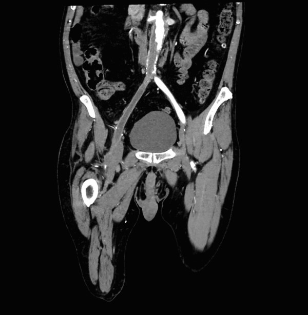
Figure 1
With a diagnosis of acute limb ischemia clinched, an argatroban drip was started. The patient crossmatched for two units of blood and was expediently transported to the operating room under the care of vascular surgery, where a right through-knee amputation was performed along with multiple embolectomies and stents.
Hospital Course
The patient was admitted to the ICU for cardiac monitoring and q30 minute right femoral pulse checks. He was successfully extubated on post-operative day 1.
On post-op day 2, however, the patient developed hypotension with an elevated jugular venous pulse to his earlobe, dependent edema, and new bibasilar crackles on lung auscultation. ECG showed new T-wave changes without ST-segment elevation, but repeat labs revealed a new serum troponin elevation — this patient was experiencing an NSTEMI. A newly elevated BNP of 6,027 prompted a transthoracic echocardiogram, which showed a left ventricular ejection fraction of 40 percent. A repeat troponin was downtrending and the patient denied chest pain or any other symptoms, so the ICU team diuresed with Lasix, made the patient NPO, and prepared him for cardiac catheterization the following morning.
Cardiac catheterization revealed chronic total occlusion of the right circumflex artery (RCA). The myocardium typically perfused by the RCA had developed retrograde collateral flow from the left anterior descending artery (LAD) to compensate; however, this patient’s new hypercoagulable state and the physiologic stress of his ischemic limb had led to severe LAD stenosis, causing his NSTEMI. Two LAD stents were placed, the patient stabilized, and he was returned to the ICU. An above-knee amputation was completed two days later (delayed due to the NSTEMI) with right groin washout and wound vac placement. The patient then underwent careful medical optimization with daily physical and occupational therapy (PT/OT). Ultimately, this patient was discharged to a nursing and rehabilitation center for continued PT/OT and daily Eliquis and Brilinta after a 19-day hospital admission.
Discussion
Acute limb ischemia (ALI) is a thromboembolic condition that occurs when arterial supply to the affected extremity becomes critically compromised, limiting perfusion and threatening limb viability if not emergently identified and managed. Thrombotic events are four times more likely to be the cause of an acutely ischemic limb than an embolic event.1 The lower extremity is 10 times more likely than the upper extremity to experience a thrombotic event, and three times more likely to experience an embolic event.4 Collateral vessels may develop in patients with underlying thrombosis; however, these collateral vessels cannot effectively perfuse the limb once complete primary vessel occlusion occurs.2 Prompt diagnosis and intervention is critical, with as many as 10 to 15 percent of patients hospitalized for acute limb ischemia ultimately requiring amputation of the affected extremity.3
ALI primarily occurs in patients with a history of peripheral vascular disease, atrial fibrillation, large vessel aneurysm, or hypercoagulable states (e.g., COVID-19).1 The classic presentation of an acutely ischemic limb includes the “6 P’s”: unilateral pain, pallor, paresthesia, paralysis, pulselessness, and poikilothermia. However, these are late findings in extremities at high risk of amputation.6 Severity is graded according to the Rutherford classification scale,1 which is used by vascular surgery to drive their management plan with regards to how salvageable the limb may or may not be. Time is tissue, as the saying goes, and prompt diagnosis is critical — irreversible necrosis begins as early as six hours after the primary vessel becomes occluded.5 While an ankle-brachial index may be performed, CT angiography is the diagnostic study of choice due to its high sensitivity and specificity.1
Clinical Pearls
Acute limb ischemia is a devastating condition if not expediently recognized and managed. While acute bullae formation are typically associated with primary infectious, dermatologic, or autoimmune etiologies (e.g., necrotizing fasciitis, SJS/TEN, bullous pemphigoid), acute limb ischemia may also present with these findings and must be considered.
Once diagnosed, anticoagulation should be initiated and vascular surgery promptly consulted for either medical or surgical management. Direct thrombin inhibitors such as argatroban should be substituted when heparin is contraindicated, such as in this patient with HIT.
It is critical to understand that a patient who is hypercoagulable enough to cause acute limb ischemia is at high risk for thromboembolic disease elsewhere, and clinicians must have a high index of suspicion for impending complications such as coronary artery disease, stroke/TIA, pulmonary embolism, etc., in these patients, as evidenced by this patient’s NSTEMI during his ICU stay.
The views expressed in this article are those of the authors and do not necessarily reflect the official policy or position of the Department of the Navy, Department of Defense, or United States Government.
References
- Mitchell ME, Carpenter JP, et al. Clinical features and diagnosis of acute lower extremity ischemia. UpToDate. Last updated: Dec 2021. Accessed: 30 Jan 2022.
- Santistevan JR. Acute Limb Ischemia: An Emergency Medicine Approach. Emerg Med Clin North Am. 2017;35(4):889-909.
- Alberino A, Eggeman D, et al. Acute arterial ischemia. WikEM. Last updated: Sep 2021. Accessed: 29 Jan 2022.
- Swaminathan, A. Vascular Disasters. Rebel EM Core Cast. Last updated: March 2020. Accessed: 29 Jan 2022.
- Simon, E. EM at 3AM — Acute Limb Ischemia. EM Docs. Last updated: March 2017. Accessed: 29 Jan 2022.
- Purcell D, Salzberg M, et al. Acute limb ischemia: Pearls and Pitfalls. EM Docs. Last updated: Feb 2015. Accessed: 29 Jan 2022.