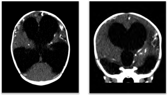
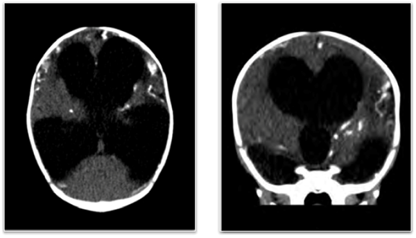
A 3-month-old female born at 37 weeks via vacuum-assisted vaginal delivery presented as a referral to the emergency department from an outpatient imaging center after obtaining a computed tomography (CT) scan of the brain to evaluate a misshapen head. On examination, she was active and had a full anterior fontanelle with a cranial deformity most prominent at the occiput, bilateral horizontal nystagmus, and globally increased muscle tone. CT brain demonstrated marked hydrocephalus with a preserved fourth ventricle, extensive cerebral atrophy, and scattered calcifications along the gray-white matter junction.
What is your diagnosis?

Congenital Toxoplasmosis
Congenital toxoplasmosis is an infection acquired in utero by transmission of the protozoan parasite Toxoplasma gondii. Transmission occurs across the placenta from mothers exposed to cat feces; those who consume raw or undercooked meats, fruits, or vegetables; or those who are immunosuppressed.1 The prevalence in the United States based on neonatal serologic screening is approximately 1 in 10,000 live births.2
The parasite causes necrosis within all parts of the central nervous system, including the cerebrum, cerebellum, brainstem, and spinal cord. Regions of necrosis often undergo calcification from an immature immune system and the resulting impaired phagocytic ability of macrophages.3
The classic triad of signs are chorioretinitis, intracranial calcifications, and hydrocephalus causing profound visual and neurodevelopmental abnormalities.1 The patient was admitted for ventriculoperitoneal shunt placement. She was started on a regimen of drugs that inhibit the synthesis of tetrahydrofolate (pyrimethamine and sulfadiazine), as well as folinic acid, which prevents bone marrow suppression from pyrimethamine.1
References
1. Jones J, Lopez A, Wilson M. Congenital toxoplasmosis. Am Fam Physician. 2003;67(10): 2131-2146.
2. Guerina NG, Hsu HW, Meissner HC, et al. Neonatal serologic screening and early treatment for congenital Toxoplasma gondii infection. N Engl J Med. 1994;330(26):1858-1863.
3. Nickerson JP, Richner B, Santy K, Lequin MH, Poretti A, Filippi CG, Huisman TA. Neuroimaging of Pediatric Intracranial Infection - Part 2: TORCH, Viral, Fungal, and Parasitic Infections. J Neuroimaging. 2012; 22(2):e52-63.


