Acute flaccid paralysis is among the most feared presentations of multiple disease processes such as Guillain-Barré syndrome (GBS) and, before vaccines, polio.
A progressively more concerning cause of acute flaccid paralysis is West Nile Virus (WNV) poliomyelitis. The incidence of this disease is increasing at an alarming rate in the United States. This devastating infectious disease affects anterior horn cells of the spinal cord, exhibiting pathophysiology that is similar to polio. Unlike polio, there is no licensed vaccine available to humans.1
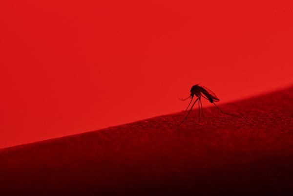
First introduced into the U.S. in 1999 during an outbreak in New York, WNV is spread via mosquitoes that transmit the virus from birds, which act as amplifying hosts. Humans and other mammals serve as “dead-end” hosts, meaning they become infected, but viruses are not transmitted between mammals. The only known disease prevention measure is to stop avian-to-human virus transmission via preventing mosquito bites and mosquito population control.
Clinically, WNV illness classically presents with fever and typical viral syndrome symptoms, such as headache, myalgia, arthralgia, nausea, vomiting, diarrhea, and/or rash. WNV also causes an alarmingly high rate of meningitis, encephalitis, and neuromuscular disease, including a polio-like paralysis, collectively termed “neuroinvasive disease.”
From 1999-2020, there were 52,000 documented cases of human WNV, and 25,000 cases of WNV neuroinvasive disease in the U.S. alone.2 Endemic to the state of Arizona, human WNV infections are rising at an unprecedented level. In 2020, there were 11 combined probable/confirmed cases of WNV reported in Arizona, and only 1 case of acute flaccid paralysis.3,4 The following year, in 2021, there were a shocking 1,644 combined probable/confirmed cases of WNV reported in Arizona. Of these cases, 1,114 were neuroinvasive, and 77 cases occurred with acute flaccid paralysis.5,6
Although Arizona is being affected at the highest rate, WNV has been detected in both humans and animals in every U.S. state. The year 2021 marked a time when human WNV neuroinvasive disease incidence peaked across the country, increasing 20%. From 1999-2019, cumulative U.S. data indicated that, on average, 49% of WNV cases were neuroinvasive. In 2021, the percentage of WNV that caused neuroinvasive disease in the U.S. increased to 69% — in just one year.2,6
This disease affects the immunocompromised and elderly the most, but all U.S. inhabitants are at risk. It is unknown which region(s) of the country will be affected next by an onslaught of neuroinvasive WNV disease. The ability to recognize, diagnose, and manage this disease is essential to the delivery of high-quality care to ED patients, many of whom (along with health care providers) are at risk for this devastating illness.
Case
In October 2021, a 50-year-old Spanish-speaking male with known uncontrolled type II diabetes, diabetic neuropathy, congestive heart failure (CHF), schizophrenia, prior transient ischemic attacks (TIA), and a history of remote traumatic brain injury (TBI) presented to an academic ED in Arizona for worsening dyspnea. He was well-known to the ED and well-documented as a difficult historian and non-compliant with medications. History-taking (with a qualified interpreter) was limited, as the patient was tangential in his responses. Although he was focused on his dyspnea, he incidentally reported a new onset of inability to ambulate, which began as lower extremity weakness approximately two months ago. When this first began, he reported to the ED with lower extremity weakness during a febrile illness, was admitted for CHF exacerbation, and was prematurely diagnosed with a viral syndrome, with diabetic polyneuropathy cited as the cause of this weakness. Despite worsening motor function, he had not pursued further work-up for this issue since that visit. There was no prior analysis of cerebrospinal fluid (CSF) or spinal cord imaging. He denied recent trauma, urinary retention, saddle anesthesia, and loss of bowel or bladder function. He denied a history of alcohol/intravenous drug abuse, recent travel, or exposure to known infectious diseases.
Physical Exam
Vital signs showed a temperature of 36.9 degrees Celsius, heart rate of 96, blood pressure 176/107, respirations 25, and oxygen saturation of 95% on room air. Physical exam demonstrated an ill-appearing man with profound lower extremity weakness and inability to ambulate. He had 2/5 strength in the bilateral lower extremities. Deep tendon reflexes (DTRs) were absent in both lower extremities. Bilateral upper extremities exhibited 2+ DTRs with 5/5 strength. His sensation was intact and symmetric throughout. Cranial Nerves II – XII were intact. No meningeal signs, facial asymmetry, dysarthria, or signs of trauma were appreciated. His neck and back were non-tender to palpation without step-offs or deformities. He was alert and oriented to person, place, time, and event. However, he was seemingly confused about the timeline of his symptoms and displayed tangential thoughts during conversation. A small maculopapular rash was noted across the anterior chest.
Studies
Chest X-ray showed cardiomegaly and bilateral pleural effusions, with a pro-Brain Natriuretic Peptide level of 831pg/mL, consistent with CHF exacerbation. His electrocardiogram did not demonstrate acute abnormalities. His troponin and complete blood cell count were within expected limits. Blood glucose level was 213 mg/dL. Otherwise, comprehensive metabolic panel was normal. A negative inspiratory force test was performed, which was within normal limits, indicating the patient had adequate diaphragm muscle tone.
Spinal magnetic resonance imaging (MRI) showed T2 hyperenhancement along the entire spinal cord, but no acute fractures or cord compression. A computed tomography scan of the patient’s brain showed encephalomalacia of the left frontal and anterior temporal pole — stable from prior studies. There was no evidence of acute infarct, hemorrhage, or enhancement to suggest meningitis.
A lumbar puncture was performed with CSF analysis. The CSF sample was clear and colorless without red blood cells. There were 27 white blood cells (WBCs)/mm3 (normal high: 5/mm3). Lymphocyte percentage was 50% and Monocytes/Macrophages percentage was 50% (reference range: 15-45%). Glucose: 61 mg/dL (within reference ranges). Protein: 93.7 mg/dL (normal high: 40 mg/dL). Oligoclonal bands were present.
Other serology tests were negative including human immunodeficiency virus screening, Lyme disease (Borrelia species), syphilis screening, tuberculosis screening, Cryptococcus antigen, and fungal serology.
Disposition and diagnosis
The patient was admitted to neurology for possible GBS or another cause of ascending paralysis and also treated for CHF exacerbation. He was discharged to a skilled nursing facility the following day with a presumptive diagnosis of paraplegia secondary to diabetic polyneuropathy.
Several days later, CSF microbiology resulted with West Nile virus IgG positive, measured at 1.59 mg/dL (reference: greater than 1.49 means antibody detected). This indicated an indolent WNV infection of the central nervous system. The WNV IgM was less than 0.90 mg/dL (antibody not detected), indicating the patient was status-post acute infection.
Other pertinent negative CSF results included a CSF gram stain, which showed white blood cells (without organisms) and cultures that showed no growth at three days. A CSF virology PCR test was negative for human herpesvirus 6, cytomegalovirus, varicella zoster virus, enterovirus, Epstein-Barr virus, herpes simplex virus (HSV) 1, and HSV 2. There was no detection of IgG or IgM to western equine encephalitis Virus, eastern equine encephalitis virus, California encephalitis virus, or St. Louis encephalitis virus.
Outcome
At the time of publication of this manuscript, the chart review indicates that this patient has been in skilled nursing facilities and never regained the ability to walk, despite rehabilitation. He has suffered from morbid sequelae of paralysis (such as sacral ulcers and osteomyelitis).
Discussion
Based on this extensive work-up, with an MRI showing T2 hyperenhancement and CSF with IgG to WNV, this patient’s diagnosis is most consistent with a subacute form of WNV poliomyelitis. A retrospective chart review revealed that the patient had presented several months ago with a febrile illness and acute onset weakness during the summer. His presentation was most consistent with acute WNV poliomyelitis at that time. This occurred during the heavy 2021 monsoon season in southern Arizona when mosquito populations flourished and the incidence of WNV cases was unprecedented.5
What is unique about this case is that the patient’s weakness was previously incorrectly diagnosed due to multiple factors that confounded his clinical presentation. Unfortunately, his symptoms were not properly worked up with spinal imaging or an LP until this ED visit. This is presumed to be due to confounding factors of medical comorbidities, priorTBI, and psychiatric diagnoses. Furthermore, severe diabetes has the propensity to worsen any neurological dysfunction, and his uncontrolled diabetes with peripheral neuropathy provided a red herring, which distracted away from his neurologic dysfunction during prior visits. Prior consultation notes by neurology indicate there was likely an anchoring bias on this aspect of his presentation. This likely caused inaccurate diagnostic momentum, which presumably caused bias in his later admitting providers. This ultimately resulted in premature closure and an incorrect diagnosis.
Any patient with encephalopathy or unexplained neurological findings in an area with mosquitos should be considered as a potential WNV neuroinvasive infection case. Neuroinvasive disease from WNV can occur with (or without) an acute febrile illness and classic symptoms of WNV. Care providers need to be aware of manifestations of WNV neuroinvasive disease, which include, but are not limited to, meningitis, encephalitis, and, with increasing incidence, neuromuscular disease, including acute flaccid paralysis.1,7
Acute onset of myalgias and weakness during infection, fever, and leukocytosis hallmark the presentation of WNV poliomyelitis. Bowel and bladder dysfunction can be seen, and encephalopathy is often present. Infrequently, numbness, paresthesia, or sensory loss can occur. The distribution of weakness commonly occurs in an asymmetric pattern, ranging from monoplegia to quadriplegia and/or diaphragm paralysis. In the absence of fever or meningoencephalitis, acute flaccid paralysis can still occur. Like polio, WNV directly affects anterior horn cells of the spinal cord and motor axons. The CSF shows pleocytosis with elevated protein levels (seen in this patient’s case). Oligoclonal bands are non-specific and, although infrequently seen, can be present in the CSF after WNV poliomyelitis (also seen in this case).1,7
Importantly, GBS and WNV poliomyelitis can be confused for one another. There are important subtle differences. In contrast, GBS demyelinates peripheral nerves and presents weeks after an acute infection, with symmetric weakness, loss of sensory function, absence of fever or leukocytosis, and with albuminocytologic dissociation in the CSF (elevated protein without pleocytosis).1,7
A neuromuscular disease differential diagnosis list should include WNV neuroinvasive disease, GBS, other immune-mediated myopathies/neuropathies, and axonal polyneuropathies (i.e., diabetic polyneuropathy). Other important considerations: bacterial meningitis or encephalitis (including Lyme disease); tick paralysis; non-infectious brain/spinal cord pathology (e.g., spinal cord compression, stroke, tumors of the central nervous system); viral meningoencephalitides (La Crosse virus, Coxsackie virus, echovirus, enterovirus); and other arbovirus encephalitides (i.e., equine encephalitis viruses, tick-borne encephalitis viruses, St. Louis encephalitis virus, and Japanese encephalitisvirus).1
To diagnose WNV poliomyelitis, LP and spinal imaging should be performed. In the setting of limb paralysis, MRI can sometimes show T2 hyperintensities, especially along the anterior horns of the spinal column (seen in this patient’s case). Serum blood tests can help elucidate the diagnosis, but analysis of CSF is the gold standard. Detection of WNV ribonucleic acid via PCR in serum and spinal fluid is possible within 3-5 days following infection but has low sensitivity compared to IgG and IgM antibodies to WNV. The IgM antibody to WNV indicates a recent infection, whereas IgG indicates a more indolent infectious process.8 Samples of CSF will generally show a moderate pleocytosis (generally greater than 200 but less than 500 WBCs/mm3), increased protein, and normal glucose.1,7
Disease prognosis varies. There is significant distress associated with early disability, but a substantial percentage of patients can recover to functional independence with intensive rehabilitation over several months. Currently, no proven effective treatment for WNV neuroinvasive disease exists. Management involves supportive care and rehabilitation. Several case series and small studies have been performed with intravenous immunoglobulin, steroids, interferon, ribavirin, and a monoclonal antibody that is no longer on the market. All therapies reported varying degrees of success rates without any reliable consistency. Ongoing research for the development of rapid antigen testing, therapies, and vaccines continues. At the time of publication, there are multiple trials still underway. There is not yet a federallyapproved, licensed vaccine available for WNV in humans. However, there are licensed vaccines for WNV available for horses. As of 2015, there have been multiple vaccine prototypes developed, some of which were reported to be in Phase 1 and Phase 2 trials, according to the National Institute of Allergies and Infectious Diseases.9,10,11
Conclusion
This case highlights key points of an uncommon disease that is re-emerging. The increasing incidence of WNV, higher rates of neuroinvasive disease, and increasing percentage of WNV poliomyelitis cases are alarming. If these trends continue, EM physicians will inevitably encounter this disease entity. It is important to consider WNV as a major cause of neuroinvasive disease and acute flaccid paralysis. If a febrile, altered patient comes into the ED with focal neurological deficits, it is vital to include this unnerving disease on the list of differential diagnoses and perform an appropriate workup. Keep in mind that WNV neuroinvasive disease can present without focal neurological deficits, and WNV poliomyelitis can occur in the absence of fever or encephalopathy. It is essential to perform the LP and order the correct diagnostic testing, including CSF antibodies to WNV (speak to your ED’s laboratory regarding various forms of WNV testing available). All providers must report positive cases of WNV to local health departments; physicians are mandated reporters. The incidence of WNV is highest in the summer months. Although we are now exiting the summer season, the incidence of WNV poliomyelitis could rise again in 2023. If this occurs, all EM physicians will also need to rise to this occasion.
Take-Home Points
• West Nile Virus (WNV) incidence has increased dramatically across the U.S. in recent years. Rates of neuroinvasive diseases caused by WNV, specifically WNV poliomyelitis, are higher than ever before.
• Know the difference between Guillain-Barré syndrome (GBS) and WNV poliomyelitis.
- WNV poliomyelitis:
- occurs during the acute phase of illness, and often presents with fever and encephalitis (but can occur without fever or encephalitis)
- usually involves asymmetric loss of motor function; the sensory function is generally preserved
- CSF shows pleocytosis with elevated protein levels
- GBS:
- occurs in the weeks after an illness/instigating event
- usually involves symmetric loss of motor and sensory function
- CSF shows albuminocytologic dissociation (elevated protein level without pleocytosis)
• Pause to consider a broad differential of etiologies when a patient has a neuromuscular disease.
• Recognize diagnostic momentum and anchoring bias as reasons for premature closure and inaccurate diagnoses, especially in complex cases with multiple confounders and social biases.
• Be a champion of public health. Scout out this disease and report it to your local health department. Physicians are mandated reporters for infectious diseases. The more public health information health departments have, the more this will help with testing, therapy, and vaccine development.
References
- Leis AA and Stokic DS. Neuromuscular manifestations of west nile virus infection. Front Neurol. 2012;3.
- Final cumulative maps and data. West nile virus. https://www.cdc.gov/westnile/statsmaps/cumMapsData.html. Published December 17, 2021. Accessed April 27, 2022.
- AZDHS: Epidemiology & disease control - mosquito borne. West nile virus - The most common mosquito borne disease in AZ. https://www.azdhs.gov/preparedness/epidemiology-disease-control/mosquito-borne/west-nile-virus/index.php. Accessed April 27, 2022.
- Arizona 2020 west nile virus statistics - azdhs.gov. https://www.azdhs.gov/documents/preparedness/epidemiology-disease-control/mosquito-borne/west-nile/data/west-nile-virus-az-2020.pdf?v=083121. Published August 31, 2021. Accessed April 27, 2022.
- Arizona 2021 west nile virus statistics - Azdhs.gov. https://www.azdhs.gov/documents/preparedness/epidemiology-disease-control/mosquito-borne/west-nile/data/west-nile-virus-stats-2021.pdf?v071321. Published March 28, 2022. Accessed April 27, 2022.
- West Nile virus disease cases by state 2021. Centers for disease control and prevention. https://www.cdc.gov/westnile/statsmaps/preliminarymapsdata2021/disease-cases-state-2021.html. Published January 11, 2022. Accessed April 27, 2022.
- Overview: West nile virus - mayocliniclabs.com. West Nile Virus Antibody, IgG and IgM, Spinal Fluid. https://www.mayocliniclabs.com/test-catalog/Overview/36772. Published 2022. Accessed April 28, 2022.
- Petersen LR, Hirsch MS, Mitty J. Clinical manifestations and diagnosis of west nile virus infection. UpToDate.https://www.uptodate.com/contents/clinical-manifestations-and-diagnosis-of-west-nile-virus-infection?search=west+nile+virus+diagnosis§ionRank=1&usage_type=default&anchor=H6&source=machineLearning&selectedTitle=1~84&display_rank=1#H6. Accessed April 28, 2022.
- West nile virus: Treatment & prevention. Centers for Disease Control and Prevention. https://www.cdc.gov/westnile/healthcareproviders/healthCareProviders-TreatmentPrevention.html#:~:text=There%20is%20no%20specific%20treatment,for%20associated%20nausea%20and%20vomiting. Published November 9, 2021. Accessed April 28, 2022.
- Santini M, Haberle S, Židovec-Lepej S, et al. Severe West Nile virus neuroinvasive disease: Clinical characteristics, short- and long-term outcomes. Pathogens. 2022;11(1):52.
- West Nile virus. National Institute of Allergy and Infectious Diseases. https://www.niaid.nih.gov/diseases-conditions/west-nile-virus. Published July 6, 2015. Accessed June 28, 2022.


