


Urachal abnormalities are a rare etiology of pediatric abdominal pain, most commonly occurring secondary to infected urachal cysts or abscesses.1 We present the case of a 10-year-old male who presented with significant suprapubic abdominal pain and was found to have an infected urachal cyst.
A 10-year-old previously healthy male presented to the pediatric emergency department with severe abdominal pain. The pain started 4 days prior and progressively worsened, with pain greatest in the suprapubic region. He developed significant dysuria, urinary frequency, and urgency over the last two days. Recent history was significant for a diagnosis of streptococcal pharyngitis two weeks prior, for which symptoms resolved and antibiotics were not initiated. The patient did not have a report of fever, nausea, vomiting, diarrhea, urethral discharge, or weight loss. The family denied a history of trauma.
On arrival in the ED, he had a temperature of 37.2 degrees Celsius, blood pressure 107/71, heart rate 104, respiratory rate 20, and oxygen saturation 99%. His exam was significant for moderate suprapubic tenderness. Obturator sign was negative, however the patient experienced significant abdominal pain with a short vertical jump. Bowel sounds were normal, and there was no distension, palpable hepatosplenomegaly, costovertebral angle tenderness, or rebound tenderness. Genitourinary exam was within normal limits, with present cremasteric reflexes, testicles in vertical lie, no discharge, and no evidence of testicular erythema, swelling, or tenderness.
The initial differential diagnosis considered included urinary tract infection, intrabdominal mass, nephrolithiasis, post-streptococcal glomerulonephritis, appendicitis, and testicular torsion. The exam was less consistent with torsion, and so serum and urine testing was sent, strep and viral swabs were obtained, and abdominal ultrasound was completed. Lab results revealed an elevated C-reactive protein 40.9 mg/L (normal <5), mildly elevated white blood cell count 11.4 k/mm3 (range 4.1-10.1) with 78% neutrophils, and mildly elevated platelets 469 k/mm3 (range 130-450). Rapid strep testing was positive. Comprehensive metabolic panel, hemoglobin, lipase, urinalysis, respiratory syncytial virus, influenza, and fecal occult blood tests were unremarkable. Abdominal ultrasound (Figure 1) was significant for a 3 cm non-compressible complex structure anterior to the bladder with echogenic and vascular structural components, and a small volume of free fluid in the lower abdomen.
Figure 1. Abdominal ultrasound image of 3 cm non-compressible complex structure, which is anterior to the bladder and posterior to the abdominal wall. This structure has anechoic and echogenic components.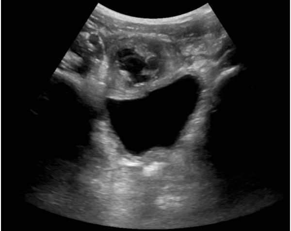
Due to the resultant consideration for malignancy as an etiology, chest x-ray was done to evaluate for associated mediastinal mass, and was negative. Given the proximity of the mass to the genitourinary system, renal ultrasound was also completed to evaluate for abnormalities, and was normal.
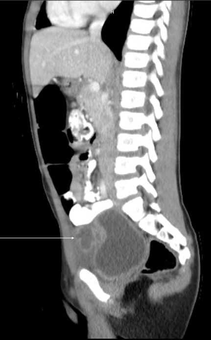
To further investigate the abdominal mass, an abdominal and pelvic computed tomography (CT) with contrast was obtained. CT demonstrated a multi-lobular collection with peripheral enhancement anterior to the bladder that measured 2.8 x 2.3 x 2.6 cm, concerning for an infected urachal cyst (Figure 2).
Figure 2. Sagittal CT image of infected urachal cyst that is anterior to bladder
Urology was consulted, and recommended admission for antibiotic coverage and pain management pending symptomatic improvement. Additionally, they recommended an outpatient urology visit within two weeks for voiding cystourethrogram (VCUG) and renal bladder ultrasound to evaluate for concomitant genitourinary pathologies and cyst resolution. Thus the patient was admitted for intravenous ceftriaxone and ketorolac.
After 3 days, the patient tolerated an oral diet with significantly improved abdominal discomfort, and he was discharged with 14 days of cefixime to continue coverage with a third generation cephalosporin. At the follow up urology appointment, VCUG was normal, while renal bladder ultrasound showed a persistent walled off lesion, and so after discussion with the family, a urachal cyst excision was scheduled.
Discussion
Urachal remnants are residual embryonic structures situated between the bladder and the umbilicus. They occur due to failed obliteration of the allantois, the structure that drains fetal urine from the bladder. This can result in a persistent patent urachus, urachal cyst, urachal sinus, or vesicourachal diverticulum.2–4 In infants, urachi typically spontaneously involute by 6 months, however this is less common in older patients.1–3 Incidence is relatively rare after infancy, with asymptomatic and symptomatic urachi detected by ultrasound in 1.6% of pediatric patients and 0.063% of adult patients.2 In patients presenting to the ED with abdominal pain, the incidence of symptomatic urachi is even lower, occurring in approximately 0.03% of abdominal pain presentations in patients aged 16 days to 59 years. 25 percent of these symptomatic urachi are in patients younger than 1 year, most commonly secondary to infected urachal cysts or abscesses.1
Urachal cysts are typically asymptomatic until infected, at which time they can present with fevers, abdominal pain, and dysuria.1,4–6 Cases can present with umbilical erythema, swelling, or drainage, though not all cases result in visible abnormalities at the umbilicus.1,4–6 On exam, tenderness is often greatest at the umbilical or suprapubic area,5–7 and a mass may be palpable at the site of maximal pain. Due to infection progression when treatment is not received, patients can also present with significant peritoneal signs and a clinical acute abdomen.7
Diagnosis of infected urachal cysts is by clinical presentation in conjunction with abdominal imaging. Imaging is most commonly initially by ultrasound, and confirmatory scans can include CT or magnetic resonance imaging for surgical management when there is a high suspicion of urachal cyst.2,4,6 Imaging of the full genitourinary tract is recommended as patients with urachal remnants may have associated genitourinary pathologies including hydronephrosis, ureteropelvic junction obstruction, and vesicoureteral reflux (VUR). Given that up to 13 percent of patients with urachal remnants have been found to have associated VUR, VCUG is a recommended component of screening.3
Broad antibiotic coverage is indicated for infected urachal cysts. This is due to the fact that several causative organisms have been cultured, including Staphylococcus aureus, Staphylococcus epidermidis, Escherichia coli, Enterococcus faecalis, Citrobacter, Proteus mirabilis, Fusobacterium, Klebsiella pneumoniae, Bacteroides, and Actinomyces israelii, with the majority of infections occuring secondary to Staphylococcus aureus.8
While surgical removal was previously recommended for all cases due to concern for future malignancy and recurrent infection, in first time presentations there is now greater consideration for conservative treatment with antibiotics and pain management. This is due to the high rate of spontaneous resolution, with up to 80% of urachal remnants resolving in infants less than 6 months old, and up to 50% resolving in childhood overall.3 Secondly, urachal malignancies cause less than 0.4% of bladder cancers, and their occurrence has not been linked to resolved pediatric urachal remnants.3 Thirdly, postoperative complication rates can reach 14.7%, with surgical complications including bladder leak or rupture, bladder diverticulum, persistent urachus, and infection.9 Therefore, conservative management is recommended as the first line approach, with surgical intervention considered for persistent urachal remnants or recurrent symptoms.3
Conclusion
Urachal cysts are embryonic remnants that can become symptomatic secondary to infection, causing suprapubic or umbilical abdominal pain. Conservative management should be highly considered in younger patients and first time presentations of urachal remnants due to high rates of spontaneous resolution.
TAKE-HOME POINTS
- Infected urachal cysts typically present with umbilical or suprapubic abdominal pain, and can have associated fevers and dysuria. Not all cases have visible umbilical abnormalities.
- Diagnosis is by clinical exam and diagnostic imaging, with ultrasound being the most common first line imaging modality.
- Given high rates of urachal remnant resolution, conservative management should be considered for first time presentations of infected urachal cysts, and the patients should follow with urology in the outpatient clinic.
References
- Schiffman JS. Urachal Remnants in Patients Presenting to the Emergency Department with Abdominal Pain. J Emerg Med. 2018;55(3):333-338. doi:10.1016/j.jemermed.2018.05.023
- Ueno T, Hashimoto H, Yokoyama H, Ito M, Kouda K, Kanamaru H. Urachal Anomalies: Ultrasonography and Management. J Pediatr Surg. 2003;38(8):1203-1207. doi:10.1016/S0022-3468(03)00268-9
- Galati V, Donovan B, Ramji F, Campbell J, Kropp BP, Frimberger D. Management of Urachal Remnants in Early Childhood. J Urol. 2008;180(4 SUPPL):1824-1827. doi:10.1016/j.juro.2008.03.105
- Yusuf S, Wassef AC, Schlesinger A. An Infected Urachal Cyst in a 4-Year-Old Girl Presenting with Recurrent Abdominal Pain. J Emerg Med. 2019;56(4):e51-e54. doi:10.1016/j.jemermed.2019.01.006
- Janes VA, Hogeman PHG, Achten NB, Tytgat SHAJ. An infected urachal cyst - A rare diagnosis in a child with acute abdominal pain. Eur J Pediatr. 2012;171(3):587-588. doi:10.1007/s00431-011-1622-3
- Gami BL, Biswas S. An infected urachal cyst. BMJ Case Rep. 2013:1-2. doi:10.1136/bcr-2012-007105
- Qureshi K, Maskell D, McMillan C, Wijewardena C. An infected urachal cyst presenting as an acute abdomen - A case report. Int J Surg Case Rep. 2013;4(7):633-635. doi:10.1016/j.ijscr.2013.02.026
- Allen JW, Song J, Velcek FT. Acute Presentation of Infected Urachal Cysts Case Report and Review of Diagnosis and Therapeutic Interventions. Pediatr Emerg Care. 2004;20(2):108-111. doi:10.1097/01.pec.0000113880.10140.19
- Naiditch JA, Radhakrishnan J, Chin AC. Current diagnosis and management of urachal remnants. J Pediatr Surg. 2013;48(10):2148-2152. doi:10.1016/j.jpedsurg.2013.02.069