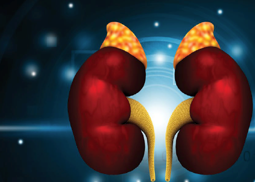
Adrenal insufficiency (AI) is a rare diagnosis in childhood and adolescence; however, it can become a major cause of morbidity and mortality in pediatric patients, as 1 in 200 cases of adrenal crisis leads to death.1
AI often presents with nonspecific, variable findings, but early recognition and treatment of these signs and symptoms can prevent detrimental outcomes. If AI goes unrecognized or treatment is delayed, children may progress to significant cardiovascular compromise and collapse.2 In the emergency department, a child may present in adrenal crisis as the initial presentation of disease or a child with a known diagnosis of primary or secondary AI may present requiring resuscitation.
In North America, the most common cause of AI is glucocorticoid withdrawal in patients on prolonged steroid therapy. Therefore, it is extremely important for primary care and emergency providers alike to recognize and take action when the condition arises.
Etiology
AI is classified as either primary (adrenal gland dysfunction) or secondary (central, hypothalamus or pituitary gland dysfunction) and includes both congenital and acquired etiologies. Causes of adrenal insufficiency in children are extensive and include enzyme deficiencies in steroidogenesis (Fig 1), metabolic and genetic disorders, autoimmune pathology, hemorrhage or infarction of the adrenal gland secondary to trauma, infection or anticoagulation, or possible drug side effects (ketoconazole, medroxyprogesterone, etomidate, rifampin, phenytoin, barbiturates).3
Congenital adrenal hyperplasia (CAH) due to 21-hydroxylase deficiency is the most common cause of primary adrenal insufficiency with an incidence of 1:10,000 to 1:15,000 people in the United States and Europe.3 All newborns in the United States are screened for 21-hydroxylase deficiency that could lead to classical CAH, a subtype of CAH that can include salt wasting and virilization, especially in female infants. Due to the absence of the 21-hydroxylase enzyme, there is a lack of cortisol, and in the case of salt-wasting CAH, a loss of aldosterone. In severe and untreated forms, this can lead to dehydration, hypotension, and shock.
Additionally, a diversion in the steroidogenesis pathway will result in androgen excess. Clinically, this can present and be diagnosed in female newborns with ambiguous genitalia at birth, whereas males will often present later at 2-3 weeks of age in a salt-wasting crisis.
The most common cause of secondary (central) AI is iatrogenic suppression of the HPA axis following prolonged use of oral glucocorticoid therapy. Long-term glucocorticoid therapy is used to treat numerous pediatric illnesses including autoimmune disease, hematological and oncological disease, inflammatory bowel disease, hematopoietic, and solid organ transplants, to name a few. Chronic glucocorticoid therapy causes suppression of ACTH, which subsequently leads to atrophy of the zona fasciculata of the adrenal cortex, the area of the adrenal gland responsible for the secretion of endogenous glucocorticoids. The duration and dosage of therapy can make a difference as well; however, there are few published studies on the duration of HPA axis suppression after glucocorticoid treatment in children, ranging from 5 days to 9 months.4 This wide variation in the timing of recovery after a course of prolonged glucocorticoid use should make providers aware of the circumstances in which AI can occur.
Diagnosis and Workup
As with any patient, paying attention to past medical history, birth history, and medications can be crucial to swift intervention for a deadly, yet oft-overlooked disease process. Adrenal crisis can be tricky to diagnose due to the nonspecific constellation of symptoms that it can present as. Additionally, the onset may be gradual. Triggers may include acute illness, physiological stress, injury, induction of anesthesia, or surgery.5
Clinically, pediatric patients may present with hypotension, tachycardia, fatigue, dizziness, nausea, vomiting, diarrhea, abdominal pain, diaphoresis, or seizures. When considering your initial differential, it is easy to assume these findings could be related to infection, other metabolic syndromes, ingestion, anaphylaxis, or a surgical process.
Initial workup should include obtaining accurate vitals, a fingerstick glucose, and a basic set of electrolytes, as well as a CBC.
In acute AI, hyponatremia is the most common lab finding. Hyperkalemia is seen in primary, but not secondary AI, and can also be accompanied by hypercalcemia and metabolic acidosis.6 Hypoglycemia is more prevalent in neonates and infants in all types of AI. Lab work may also show normocytic anemia, lymphocytosis and eosinophilia. Initial screening for primary AI can be done with a low cortisol level, <140 nmol/L (5 mcg/dL), and a plasma ACTH level greater than two-fold the upper limit of normal for the reference interval. Additionally, according to clinical practice guidelines, the gold standard of diagnostic testing with the cosyntropin test can be done to rule out primary AI in patients with unexplained symptoms as described. However, do not delay the initiation of treatment while awaiting results for cosyntropin testing. This can also be done as a confirmatory test after treatment and patient stabilization.6
Treatment in the ED
As always, first assess your patient’s ABCs, and establish IV access as soon as possible, with lab workup as discussed. However, if the blood draw is proving to be difficult, do not delay treatment.
The initial stress dose of 100 mg/2mL hydrocortisone sodium succinate should 50 mg/m^2 or if BSA is not available, can also be based on the child’s age: children ≤3 years: 25 mg; >3 and <12 years: 50 mg; and older children and adolescents ≥12 years: 100 mg as an initial stress dose. This should be followed by 50–100 mg/m2/day divided into 4 doses given every 6 hours, or as a continuous IV infusion.7,8 If hydrocortisone is not available, you can use prednisolone as an alternative.
Prompt fluid resuscitation for hypovolemic shock should be administered with 20 ml/kg boluses of isotonic fluid such as 0.9% normal saline. Additionally, hypoglycemia should be treated with an initial bolus 2-4 mL/kg D25W infused slowly at 2-3 mL/min or 5-10 ml/kg D10W IV and repeated as necessary. Start 1.5-2x maintenance fluids with D5NS.
For hyperkalemia, always be sure to obtain an EKG to check for peaked T-waves. These can progress to a prolonged PR interval or QRS duration. Usually, hyperkalemia improves with steroid and fluid therapy and rarely requires administration of insulin and glucose. Be sure to monitor and treat any other electrolyte abnormalities.
When treating an adrenal crisis, don’t forget to consider the trigger. If infection or sepsis is suspected, make sure to add on systemic antibiotics accordingly.
Once the patient is stabilized, pediatric endocrinology should then be contacted and the patient should be admitted to ICU level care with continuous hemodynamic monitoring.
Children with known AI should be on maintenance glucocorticoid replacement therapy managed by their pediatrician or endocrinologist in order to help prevent acute AI. Families should also be provided injectable intramuscular hydrocortisone sodium succinate to use at home in the event of stress/illness with vomiting or altered mental status and instructions on when to seek emergent medical care. It is recommended that primary providers also give patients an ED letter, steroid card with appropriate stress dosing, or medical alert bracelet in order to inform EMS or ED providers about their diagnosis and how to quickly initiate treatment for adrenal crisis. These steps can greatly reduce morbidity and mortality.
Conclusion
Adrenal insufficiency in the pediatric population may stem from a myriad of causes; however, what is of utmost importance is to consider acute and life-threatening adrenal crisis. This requires timely recognition and initiation of appropriate medical management with steroid therapy in order to avoid further decompensation. ED providers should be aware of this diagnosis and have a high index of suspicion for AI in critically ill patients in the setting of electrolyte abnormalities.
References
- Miller BS, Spencer SP, Geffner ME, et al. Emergency management of adrenal insufficiency in children: advocating for treatment options in outpatient and field settings. Journal of Investigative Medicine. 2019;68(1):16-25. doi:10.1136/jim-2019-000999
- Shulman DI, Palmert MR, Kemp SF. Adrenal Insufficiency: Still a Cause of Morbidity and Death in Childhood. PEDIATRICS. 2007;119(2):e484-e494.
- Speiser PW. Congenital Adrenal Hyperplasia. NORD (National Organization for Rare Disorders). https://rarediseases.org/rare-diseases/congenital-adrenal-hyperplasia/. Published June 26, 2018. Accessed April 5, 2021.
- Mendoza-Cruz AC, Wargon O, Adams S, Tran H, Verge CF. Hypothalamic-Pituitary-Adrenal Axis Recovery Following Prolonged Prednisolone Therapy in Infants. J Clin Endocrinol Metab. 2013;98(12):E1936-E1940.
- Bornstein SR, Allolio B, Arlt W, et al. Diagnosis and Treatment of Primary Adrenal Insufficiency: An Endocrine Society Clinical Practice Guideline. J Clin Endocrinol Metab. 2016;101(2):364-389.
- Cortet C, Barat P, Zenaty D, Guignat L, Chanson P. Group 5: Acute adrenal insufficiency in adults and pediatric patients. Annales d'Endocrinologie. 2017;78(6):535-543.


