


We present an uncommon case of a total knee arthroplasty (TKA) dislocation.
Knee dislocations are rare, representing a mere 0.02-0.2% of orthopedic injuries in the general population.1,2 In the even smaller subset of patients with TKAs, the risk of knee dislocation is 0.15–0.5%.3 As uncommon as these injuries are, they constitute a true emergency; what may appear at first glance to be a simple joint dislocation could be hiding more serious neurovascular damage.
Case
A 72-year-old male with a history of autoimmune hepatitis status post-transplant and, nine years earlier, a left TKA, was brought in by EMS to the emergency department shortly after a mechanical trip and fall. The patient stated that while taking out the garbage, he fell and landed on his left leg. He experienced no pain but was then unable to stand. On exam, the patient was hemodynamically stable. His left knee was immobilized in a splint from EMS and demonstrated gross deformity with a positive sag sign (Image 1). There were no breaks in the skin. Distal pulses and sensation were intact, and the patient was able to actively plantar and dorsiflex.
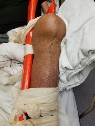
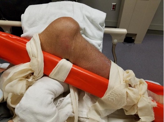
Image 1. AP (top) and lateral (bottom) views of dislocated knee in splint.
Differential diagnosis included dislocated TKA, popliteal artery or vein injury, tibial or common fibular nerve injury, or fracture of the femur, tibia, or fibula. Lateral radiographs of the left knee (Image 2) revealed a TKA with posterior displacement of the tibial component relative to the femoral component. There were no obvious fractures, but the X-ray did show non-specific thin lucency at the bone cement interface of the tibial component that could indicate loosening or infection.
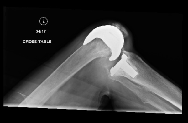
Image 2. X-ray of posterior dislocated TKA.
After providing fentanyl for analgesia, the patient’s knee was reduced with simple traction. Post-reduction physical exam showed intact distal pulses, and X-rays showed a successful reduction. Post-reduction CT angiography with runoff showed a widely patent arterial system. Orthopedic surgery assessed and cleared the patient for discharge from ED in a bulky Jones dressing and knee immobilizer. After 4 weeks, knee range-of-motion was 0 to 120 degrees with good stability and no effusion. His knee did not require revision surgery.
Discussion
Traditionally, knee dislocations are associated with high-energy trauma, but certain risk factors such as obesity have increased the prevalence of low-velocity impacts. Anterior dislocations are often caused by knee hyperextension, while posterior dislocations are classically caused by dashboard injuries or other forced posterior tibial translation in relation to the femur.1 For native knees, anterior dislocations are more common than posterior, whereas for TKAs the opposite is true.1,4 The causes of TKA dislocations are usually due to underlying issues such as implant malpositioning, soft tissue deformity, inappropriate selection of the primary implant, rupture of the PCL, polyethylene wear, and other hardware malfunction.3-5
On arrival to the ED, Advanced Trauma Life Support (ATLS) should be initiated to identify all injuries. Subsequently, the dislocated knee should be examined for deformity and neurovascular compromise. X-rays are the initial image of choice which, if possible in a rapid and timely manner, should still be performed before reduction even in a hypoperfusing limb, as they can confirm the direction of the dislocation and reveal any concomitant fractures.1,2Traction and countertraction with sedation or heavy analgesia is usually the preferred method of reduction in dislocated knees, followed by a full neurovascular exam, assessment of the knee ligaments, and immobilization of the knee in full extension.2,4 Post-reduction frequently requires CT or MR angiography, though X-rays can suffice with a normal physical exam. Neurovascularly compromised or extremely unstable knees require surgical intervention.2
Knee dislocations often self-reduce spontaneously in the field, and by the time the patient reaches the emergency department, the physical exam may at first glance appear to be normal1,2,6 — thus reinforcing the importance of a detailed neurovascular exam, including checking limb sensation, movement, perfusion, pulses, and ankle brachial pressure index.1,2,6
Both native knee dislocations and TKA dislocations may be associated with ligamentous injuries, some of which require immediate surgery and some that require surgery 2-3 weeks after the initial injury, once the inflammation has improved.2 Many patients can be managed non-operatively and must follow a months-long physical therapy and rehabilitation program before they can return to full activity.
Key Points
- Knee dislocations, whether native or TKA, are a true emergency and require immediate X-rays and reduction.
- A thorough post-reduction physical exam, including an ankle brachial pressure index, can help inform the need for further imaging with CT or MR angiography or immediate surgical intervention.
- After reduction, the neurovascularly intact knee should be immobilized in full extension until outpatient follow-up.
- Even with non-operative management, a return to full activity takes months of physical therapy and rehabilitation.
References
- McKee L, Ibrahim MS, Lawrence T, Pengas IP, Khan WS. Current concepts in acute knee dislocation: the missed diagnosis? Open Orthop J. 2014;8162-167.
- Pardiwala DN, Rao NN, Anand K, Raut A. Knee Dislocations in Sports Injuries. Indian J Orthop. 2017;51(5):552-562.
- Villanueva M, Rios-Luna A, Pereiro J, Fahandez-Saddi H, Perez-Caballer A. Dislocation following total knee arthroplasty: A report of six cases. Indian J Orthop. 2010;44(4):438-443.
- Conti A, Camarda L, Mannino S, Milici L, D’Arienzo M. Anterior dislocation in a total knee arthroplasty: A case report and literature review. J Orthop. 2014;12(Suppl 1):S130-132.
- Rouquette L, Erivan R, Pereira B, Boisgard S, Descamps S, Villatte G. Tibiofemoral dislocation after primary total knee arthroplasty: a systematic review. Int Orthop. 2019;43(7):1599-1609.
- Gottlieb M, Koyfman A, Long B. Evaluation and Management of Knee Dislocation in the Emergency Department. J Emerg Med. 2019;S0736-4679(19)30826-1.